Japanese
English
- 有料閲覧
- Abstract 文献概要
- 1ページ目 Look Inside
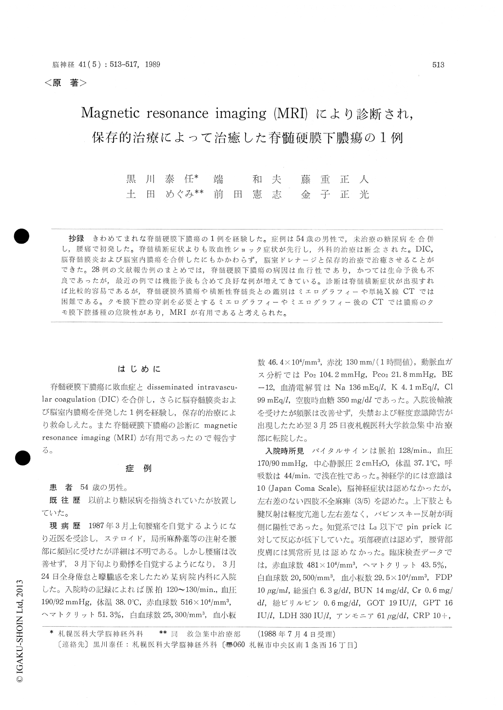
抄録 きわめてまれな脊髄硬膜下膿瘍の1例を経験した。症例は54歳の男性で,未治療の糖尿病を合併し,腰痛で初発した。脊髄横断症状よりも敗血性ショック症状が先行し,外科的治療は断念された。DIC,脳脊髄膜炎および脳室内膿瘍を合併したにもかかわらず,脳室ドレナージと保存的治療で治癒させることができた。28例の文献報告例のまとめでは,脊髄硬膜下膿瘍の病因は血行性であり,かっては生命予後も不良であったが,最近の例では機能予後も含めて良好な例が増えてきている。診断は脊髄横断症状が出現すれば比較的容易であるが,脊髄硬膜外膿瘍や横断性脊髄炎との鑑別はミエログラフィーや単純X線CTでは困難である。クモ膜下腔の穿刺を必要とするミエログラフィーやミエログラフィー後のCTでは膿瘍のクモ膜下腔播種の危険性があり,MRIが有用であると考えられた。
Localized suppuration involving the spinal cord is uncommon. A case of spinal subdural empyema is reported. The patient is 54-year-old male who had been suffering a diabetes mellitus but did not receive any treatment. His initial symptom was lumbago. Then he noticed a palpitation and gen-eral malaise which made him visit a hospital. Because he did not show any improvement by a fluid therapy, he was transferred to our institute for the further evaluation. On admission, physicalexamination showed no abnormality. Blood pres-sure was 170/90 mmHg, heart rate 128/min. and body temperature 37.1℃ suggesting a septic shock state. Neurological examination revealed slight consciousness disturbance, mild tetraparesis and bilateral hypesthesia lower than the level of L3. Laboratory examination showed the elevated leu-kocyte count and fasting blood sugar and urine keton body levels of 20, 500/mm3, 257 mg/dl and 226 mg/dl respectively. Blood culture proved a septi-cemia of Streptococcus agalactiae afterwards. On the second day of admission, lumbar puncture re-vealed a purulent cerebrospinal fluid, though X-ray CT of lumbar spine did not confirm a diagnosis. Spinal magnetic resonance imaging (MRI) revealed a widespread abnormal intensity of the spinal canal from the level of Th11 to L4. On the T1-weighted image (TR 300 msec., TE 40 msec.), cerebrospinal fluid space was abnormally isointense. On the T1-weighted image (TR 2, 000 msec., TE 80 msec.), subdural and cerebrospinal space was filled with an abnormal high-intense lesion espe-cially on the ventral side. He developed semicoma due to hydrocephalus following a intraventricular empyema. He was also complicated disseminated intravascular coagulation. His general condition forced us to do a conservative treatment of sys-temic and intraventricular administration of anti-biotics combined with a continuous ventricular drainage. Although he did not receive a surgical treatment against spinal subdural empyema, he was recovered and became ambulant.
Twenty-eight cases of spinal subdural empyema have been reported. Some previous cases showed a poor life prognosis, though they received a surgical treatment of spinal subdural empyema. Recently cases with good functional recovery have been re-ported with or without surgical treatment.
The accurate diagnosis of spinal subdural empye-ma is not always easy. Since spinal subdural em-pyema does not have a characteristic clinical fea-ture, it sometimes should be differentiated from other spinal lesions such as epidural empyema and transverse myelitis. Conventional methods for definite diagnosis of the lesion require a cerebro-spinal fluid space puncture which would cause a dissemination of the empyema to the whole central nervous system. MRI was most useful for the di-agnosis of this rare condition, while X-ray CT was not helpful as shown in our case.
It should be noticed that MRI would contribute to the definite diagnosis of subdural empyema and make it possible to perform a prompt surgical treatment unless severe spinal damage occurs.

Copyright © 1989, Igaku-Shoin Ltd. All rights reserved.


