Japanese
English
- 有料閲覧
- Abstract 文献概要
- 1ページ目 Look Inside
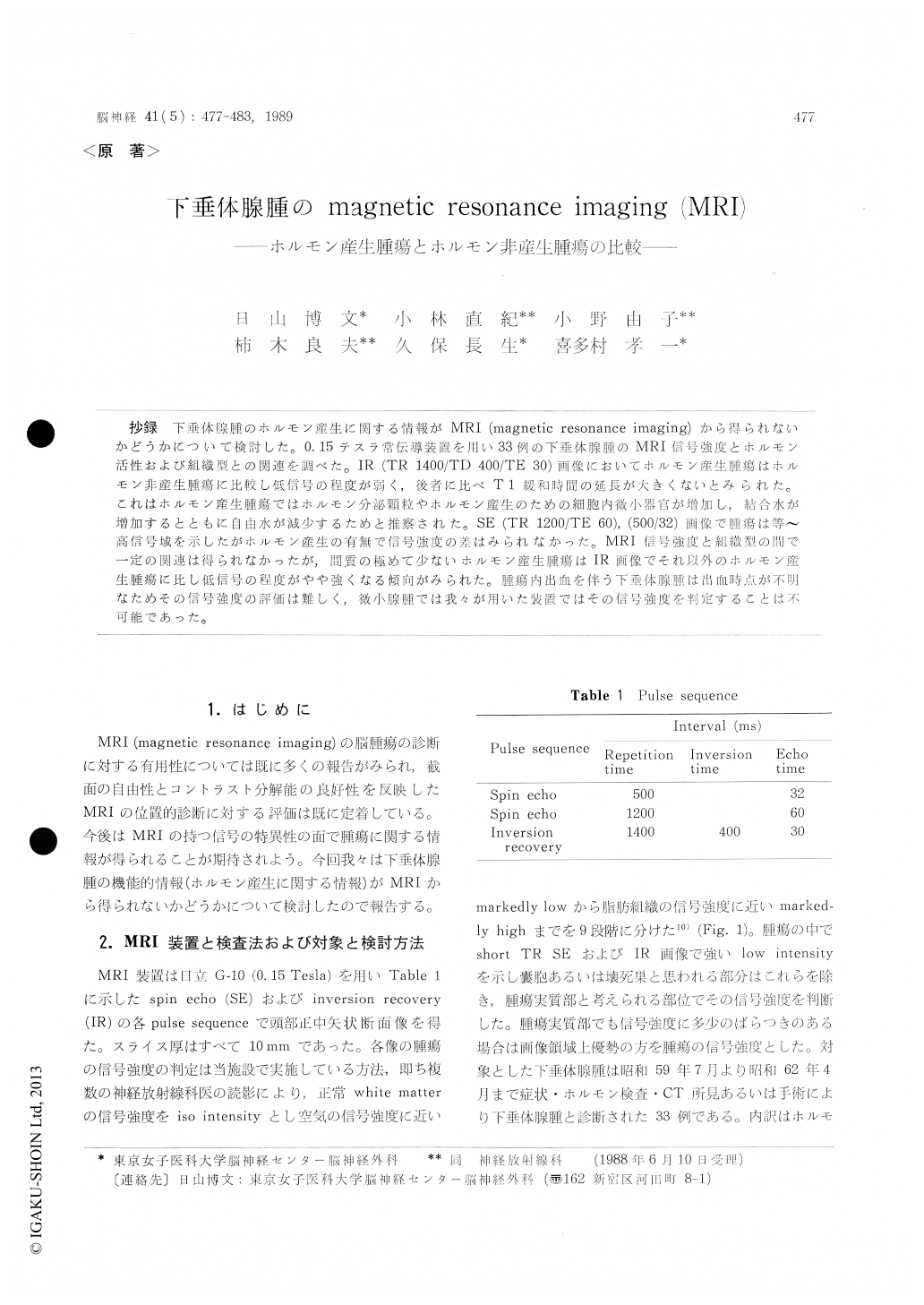
抄録 下垂体腺腫のホルモン産生に関する情報がMRI (magnetic resonance imaging)から得られないかどうかについて検討した。0.15テスラ常伝導装置を用い33例の下垂体腺腫のMRI信号強度とホルモン活性および組織型との関連を調べた。IR (TR 1400/TD 400/TE 30)画像においてホルモン産生腫瘍はホルモン非産生腫瘍に比較し低信号の程度が弱く,後者に比べT1緩和時間の延長が大きくないとみられた。これはホルモン産生腫瘍ではホルモン分泌顆粒やホルモン産生のための細胞内微小器官が増加し,結合水が増加するとともに自由水が減少するためと推察された。SE (TR 1200/TE 60), (500/32)画像で腫瘍は等〜高信号域を示したがホルモン産生の有無で信号強度の差はみられなかった。MRI信号強度と組織型の間で一定の関連は得られなかったが,間質の極めて少ないホルモン産生腫瘍はIR画像でそれ以外のホルモン産生腫瘍に比し低信号の程度がやや強くなる傾向がみられた。腫瘍内出血を伴う下垂体腺腫は出血時点が不明なためその信号強度の評価は難しく,微小腺腫では我々が用いた装置ではその信号強度を判定することは不可能であった。
Thrity three cases of pituitary ademoma were examined by MRI with 0.15 T system. Eight cases of functioning tumor showed iso---minimally low intensity, and 10 cases of non-functioning tu-mor did slightly--,markedly low intensity on IR image. Functioning tumor cells contain well-developed rough-surfaced endoplasmic reticulum, Golgi complexes and numerous secretary granule, so that bound water more increases and T 1 re-laxation time less prolongs in functioning tumor than in non-functioning tumor. Two cases of func-tioning tumor disclosed slightly low intensity on IR image because of its poor stroma. It is neces-sary to know the exact bleeding time so as to measure the signal intensity in pituitary apoplexy case. Microadenoma appeared spotty hypointensity and upward convexity of superior surface of the gland on IR image.

Copyright © 1989, Igaku-Shoin Ltd. All rights reserved.


