Japanese
English
- 有料閲覧
- Abstract 文献概要
- 1ページ目 Look Inside
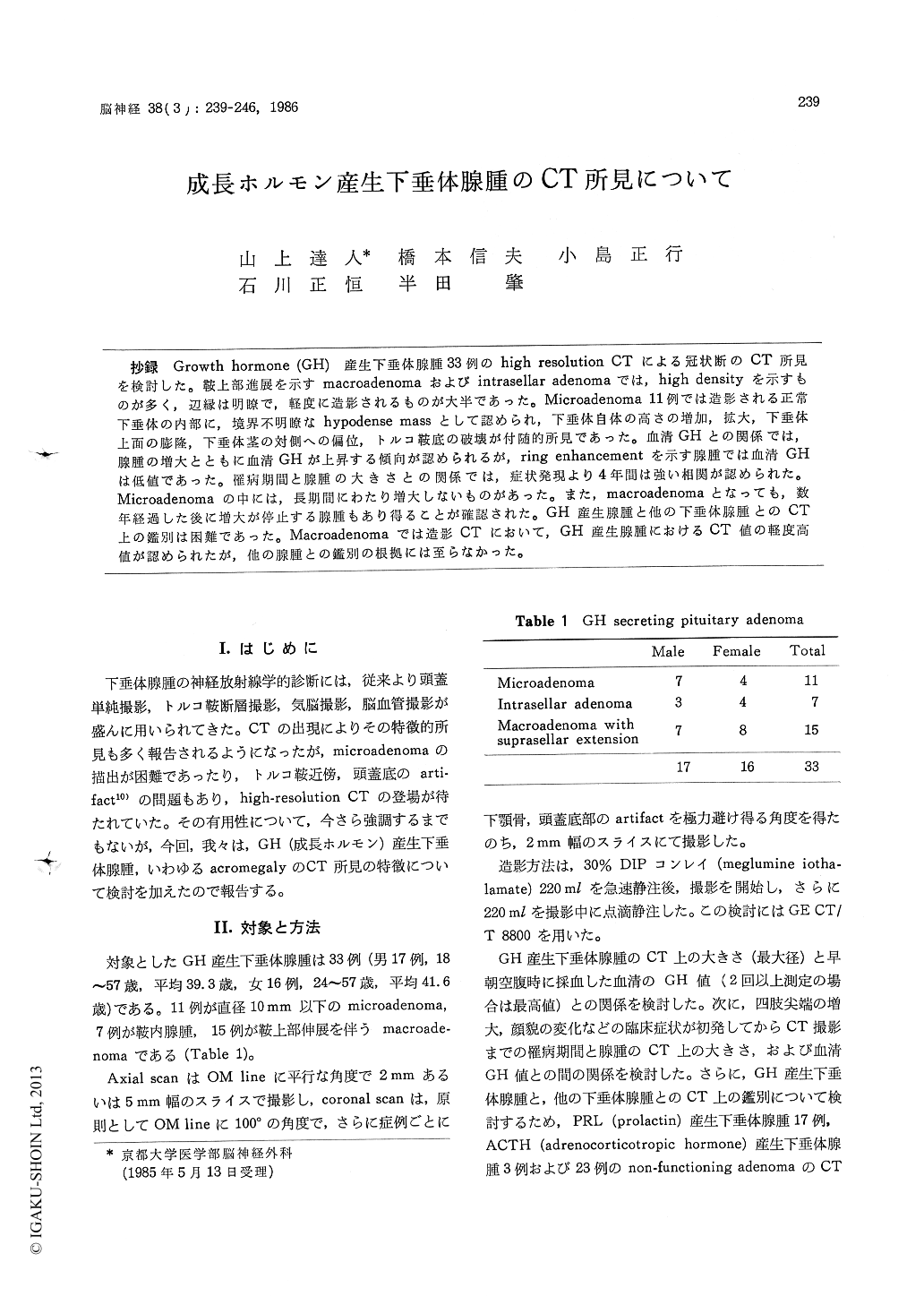
抄録 Growth hormone (GH)産生下垂体腺腫33例のhigh resolution CTによる冠状断のCT所見を検討した。鞍上部進展を示すmacroadenomaおよびintrasellar adenomaでは, high densityを示すものが多く,辺縁は明瞭で,軽度に造影されるものが大半であった。Microadenoma 11例では造影される正常下垂体の内部に,境界不明瞭なbypodense massとして認められ,下垂体自体の高さの増加,拡大,下垂体上面の膨隆,下垂体茎の対側への偏位,トルコ鞍底の破壊が付随的所見であった。血清GHとの関係では,腺腫の増大とともに血清GHが上昇する傾向が認められるが, ring enhancementを示す腺腫では血清GHは低値であった。罹病期間と腺腫の大きさとの関係では,症状発現より4年間は強い相関が認められた。Microadenomaの中には,長期間にわたり増大しないものがあった。また, macroadenomaとなっても,数年経過した後に増大が停止する腺腫もあり得ることが確認された。GH産生腺腫と他の下垂体腺腫とのCT上の鑑別は困難であった。Macroadenomaでは造影CTにおいて, GH産生腺腫におけるCT値の軽度高値が認められたが,他の腺腫との鑑別の根拠には至らなかった。
The value of high-resolution computed tomo-graphy (CT) in the diagnosis of pituitary adenoma has recently been stressed, especially of the coro-nal view with contrast enhancement. Analysis of the CT scans of 33 growth hormone (GH) secre-ting pituitary adenomas was done (11 cases of microadenomas, 7 cases of intrasellar adenomas and 15 cases of macroadenomas with suprasellar extension).
In macroadenomas, the density was high in five cases, high with isodense portion in two cases, mixed in four cases, isodense in three cases, and isodense and low dense in one case. Six adenomas showed homogeneous density and nine were hetero-geneous. After contrast enhancement, two cases showed marked enhancement, ten cases mild and three cases ring enhancement. Margin of adenoma was smooth in nine cases and irregular in six. Among seven cases of intrasellar adenoma one accompanied primary empty sella.
In microadenomas ten of eleven cases had hypo-dense mass inside the normally enhanced pituitary gland. The margin was ill-defined in seven cases and well-defined in three. Eight cases had pituitary height 7 mm or more. Upper surface of the pitui-tary gland was convex upward in five cases, flat in four and concave in two. Deviation of pituitary stalk was found in seven cases. Bony changes of sellar floor were recognized in three cases. There was a tendency that serum GH level increased with the increment of the size of ade-noma. Serum GH levels in adenomas with ring enhancement were lower than those in the homo-geneously enhanced adenomas of similar size. One case with marked enhancement showed the highest GH level among all adenomas of the presented series.
There was a positive correlation between the size of GH secreting adenoma and the length of clinical history, especially during the early four years in the course of the disease. Some micro-adenomas with long clinical histories indicate that there are some adenomas which do not grow in size for a long time.
The CT number (Hounsfield Unit) of the GH secreting adenoma was from 40 to 49 in plain scan and from 56 to 99 in enhanced scan. Although CT number of GH secreting adenomas was higher than those of other types of adenoma, the dif-ference was not significant as to differentiate the types of adenoma.

Copyright © 1986, Igaku-Shoin Ltd. All rights reserved.


