Japanese
English
- 有料閲覧
- Abstract 文献概要
- 1ページ目 Look Inside
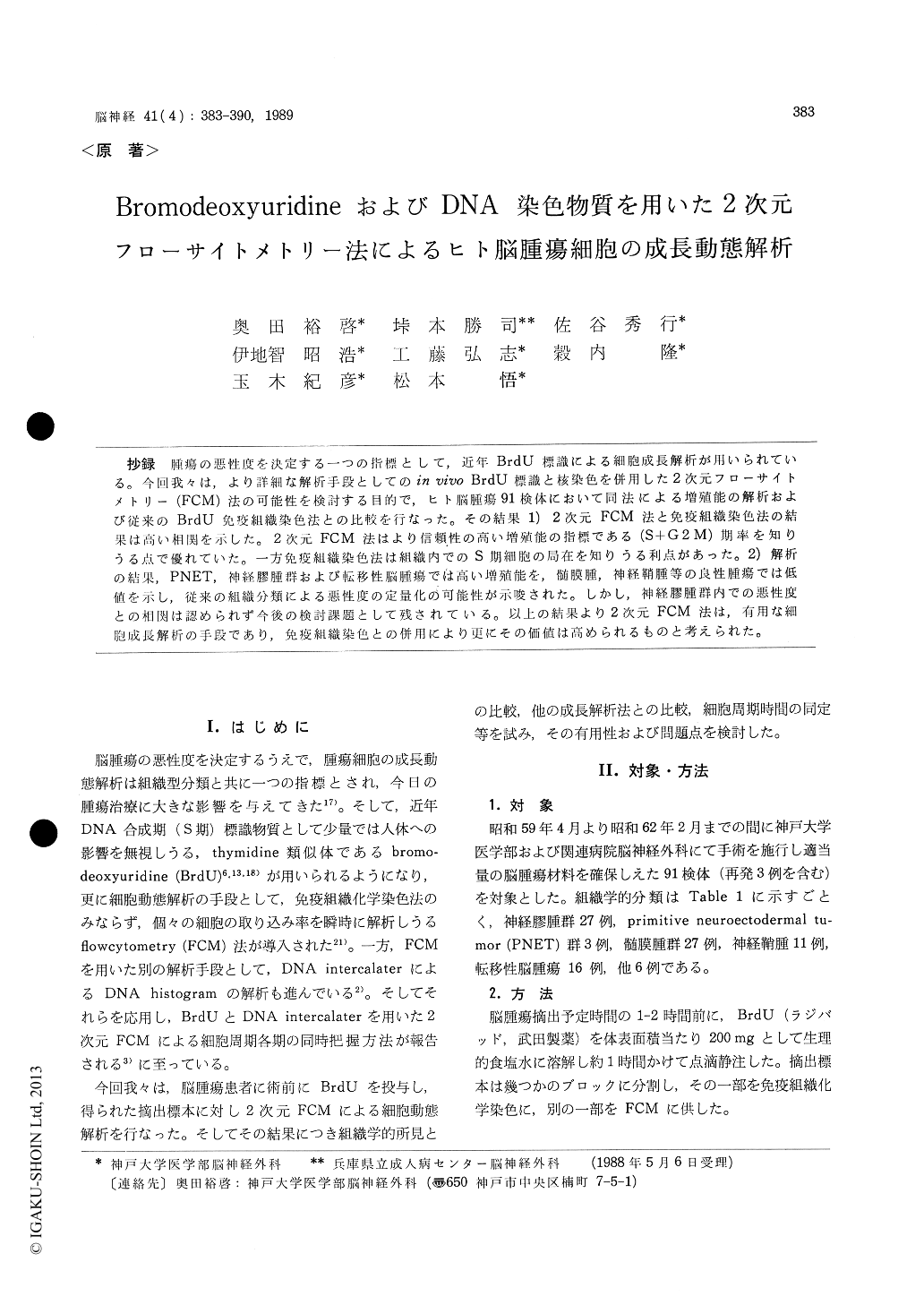
抄録 腫瘍の悪性度を決定する一つの指標として,近年BrdU標識による細胞成長解析が用いられている。今回我々は,より詳細な解析手段としてのin vivo BrdU標識と核染色を併用した2次元フローサイトメトリー(FCM)法の可能性を検討する目的で,ヒト脳腫瘍91検体において同法による増殖能の解析および従来のBrdU免疫組織染色法との比較を行なった。その結果1)2次元FCM法と免疫組織染色法の結果は高い相関を示した。2次元FCM法はより信煩性の高い増殖能の指標である(S+G2M)期率を知りうる点で優れていた。一方免疫組織染色法は組織内でのS期細胞の局在を知りうる利点があった。2)解析の結果,PNET,神経膠腫群および転移性脳腫瘍では高い増殖能を,髄膜腫,神経鞘腫等の良性腫瘍では低値を示し,従来の組織分類による悪性度の定量化の可能性が示唆された。しかし,神経膠腫群内での悪性度との相関は認められず今後の検討課題として残されている。以上の結果より2次元FCM法は,有用な細胞成長解析の手段であり,免疫組織染色との併用により更にその価値は高められるものと考えられた。
Cell kinetics of 91 human brain tumors obtained from 88 patients were analyzed with the following two methods, 1) bivariate (two-color) flowcyto-metric measurement of cellular DNA content and amount of bromodeoxyuridine (BrdU) incorporated into cellular DNA, in 66 specimens, 2) immunohis-tochemical detection of BrdU incorporated S-phase cells, in 34 specimens.
Patients were given an intravenous 1 hour in-fusion of 200 mg/sq. m. of BrdU 1-2 hours before the surgical removal. The excised tumor specimen was divided into several portions. One was fixed with 70% ethanol and embedded in paraffin, and another was digested mechanically and/or chemi-cally to obtian a single cell suspension, and fixed in 70% ethanol. Paraffin-embedded tissue sections were stained by the peroxidase-antiperoxidase im-munohistochemical method using anti-BrdU mono-clonal antibody (MoAb). Single cell suspensions were reacted with fluorescein isothiocyanate (FITC) conjugated anti-BrdU MoAb, or anti-BrdU MoAb and FITC-conjugated second antibody suc-cessively by the staining with propidium iodide, for flowcytometry (FCM).
Rates of S-phase fraction in single cell suspen-sions calculated by bivariate FCM were correlated well with labeling indexes (LI, i.e. the percentage of BrdU incorporated cells) calculated in tissue sections, but not with the result of analysis of DNA histogram by Dean's method. This discrepan-cy is probably due to large coefficient value in several samples. Histological malignancy of the tumors was reflected both in the proliferating index (PI, i. e. % S+G2M phase) calculated by bivariate FCM and the LI by immunohistochemical method. PI tended to be high in primitive neuro-ectodermal tumors and metastatic carcinomas, moderately high in gliomas, and low in benign tumor groups. LI in immunohistochemical studies showed the same tendency.
The advantages of bivariate FCM method were 1) able to simultaneously detect each phase (G0+ Gl, S and G2M) of the cell cycle, and polyploidy, 2) provide accurate information on over 10000 individual cells in a short time. On the other hand, immunohistochemical staining provided informa-tion about the in situ relationship between the histological malignancy and the location of BrdU incorporated cell clusters. These results indicate that the bivariate FCM analysis of tumor cell kine-tics may be useful in estimating the biological malignancy, especially in combination with the immunohistochemical staining of BrdU incorpo-rated cells.

Copyright © 1989, Igaku-Shoin Ltd. All rights reserved.


