Japanese
English
- 有料閲覧
PPV購入案内
1,320円 (1,200円+税10%) こちらは、27ページから35ページの文献です。購入には医書.jp本会員登録が必要です。
会員登録完了後に改めて上記「購入サイトへ」を選択してください。
または「購入サイトへ」選択後に「購入ログイン」、「新規会員登録」の順に進んでください。
会員登録について詳しくはをご確認ください。
- Abstract 文献概要
- 1ページ目 Look Inside
I.はじめに
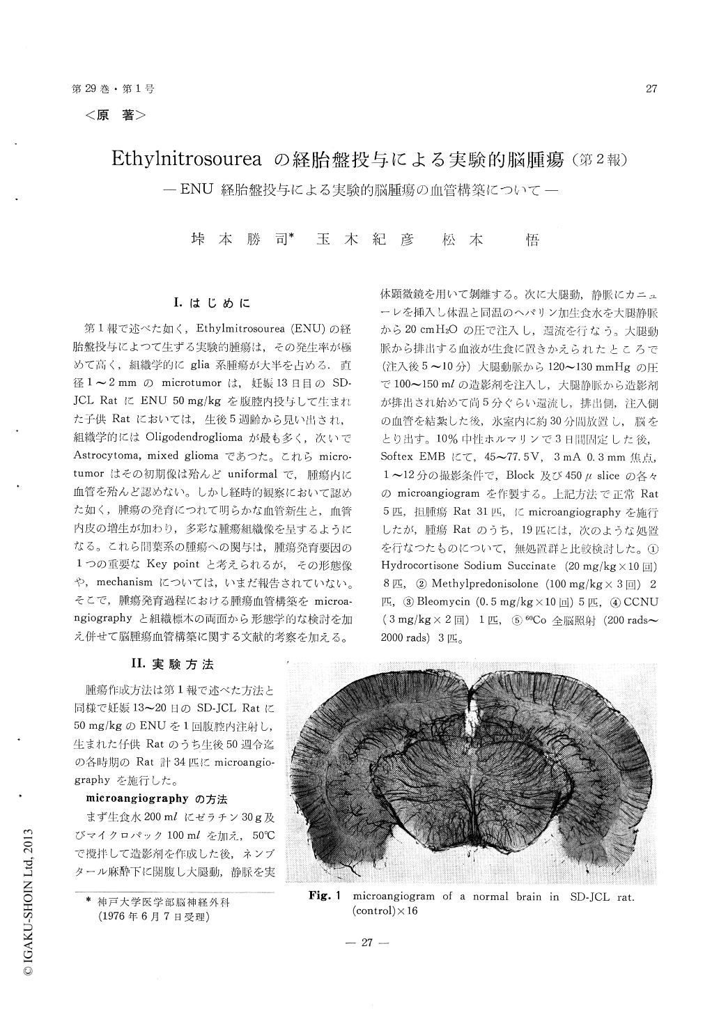
第1報で述べた如く,Ethylmitrosourea (ENU)の経胎盤投与によつて生ずる実験的腫瘍は,その発生率が極めて高く,組織学的にglia系腫瘍が大半を占める.直径1〜2mmのmicrotumorは,妊娠13日目のSD—JCL RatにENU 50mg/kgを腹腔内投与して生まれた子供Ratにおいては,生後5週齢から見い出され,組織学的にはOligodendrogliomaが最も多く,次いでAstrocytoma, mixed gliomaであつた。これらmicro—tumorはその初期像は殆んどuniformalで,腫瘍内に血管を殆んど認めない。しかし経時的観察において認めた如く,腫瘍の発育につれて明らかな血管新生と,血管内皮の増生が加わり,多彩な腫瘍組織像を呈するようになる。これら間葉系の腫瘍への関与は,腫瘍発育要因の1つの重要なKey pointと考えられるが,その形態像や,mechanismについては,いまだ報告されていない。そこで,腫瘍発育過程における腫瘍血管構築をmicroa—ngiographyと組織標本の両面から形態学的な検討を加え併せて脳腫瘍血管構築に関する文献的考察を加える。
The architecture of the vessels of the experi-mental brain tumors produced transplacentally by Ethylnitrosourea (ENU) in SD-JCL rats was in-vestigated by microangiography, as compared with those of normal rat brains.
In a normal brain, cortical vessels were distri-buted evenly throughout the brain surface and extended into the white matter in parallel. In microangiography at the stage of the microtumor, no remarkable change was found in the blockspecimen of the brain, but very fine abnormal vessels with surrounding poor vascularity could be seen in the white matter of a 2 mm-sliced specimen of the brain.
In the middle sized tumor, extravasations of the contrast media and microaneurysms were found in the center of the tumor. The cortical vessels were disordered, and a part of these vessels increased in number and extended into the tumor region. Ir-regular, spiral, and tortuous was each capillary that was different from the normal vessels in the respect of the course, arrangement, and morphology.
As it was growing, the tumor had many ab-normal vessels occupying the greater part of the hemisphere. In addition, small multifocal extra-vasations of the contrast media was noticed in the tumor, and tumor vessels were very irregular, tortous, dilated, and kinked. Angiomatous changes,reticular formation of the vessels, and glomerular formation of the capillaries, namely the proliferation of the endothelial cells, were also found around the marginal zone of the tumor.
In a large tumor, normal cortical vessels had been lost and the necrosis or pooling of the contrast medium was noticed in the center of the tumor. The vessels around the marginal zone of the tumor were kinking and some of them stretched. The active growth of the tumor was indicated by the marked proliferation of the tumor vessels.
Some changes of the tumor vessels such as small bleeding and slight proliferation of the capillary endothelium were seen in the group of 60Co irra-diation as well as that of combined administration of the Steroids and Bleomycin.
Copyright © 1977, Igaku-Shoin Ltd. All rights reserved.