Japanese
English
- 有料閲覧
- Abstract 文献概要
- 1ページ目 Look Inside
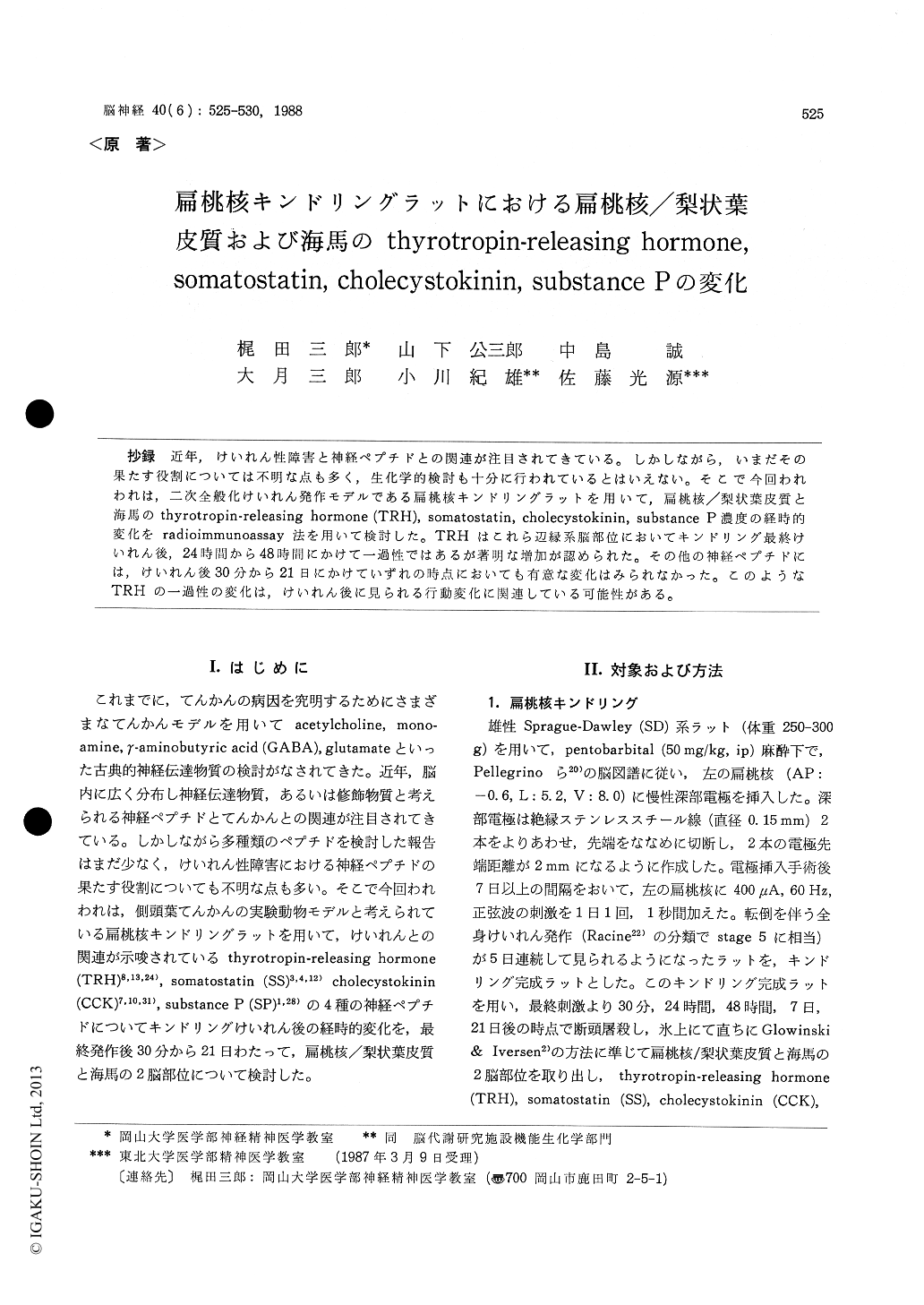
抄録 近年,けいれん性障害と神経ペプチドとの関連が注目されてきている。しかしながら,いまだその果たす役割については不明な点も多く,生化学的検討も十分に行われているとはいえない。そこで今回われわれは,二次全般化けいれん発作モデルである扁桃核キンドリングラットを用いて,扁桃核/梨状葉皮質と海馬のthyrotropin-releasing hormone (TRH), somatostatin, cholecystokinin, substance P濃度の経時的変化をradioimmunoassay法を用いて検討した。TRHはこれら辺縁系脳部位においてキンドリング最終けいれん後,24時間から48時間にかけて一過性ではあるが著明な増加が認められた。その他の神経ペプチドには,けいれん後30分から21日にかけていずれの時点においても有意な変化はみられなかった。このようなTRHの一過性の変化は,けいれん後に見られる行動変化に関連している可能性がある。
Recently, several systems of neuropeptides have been demonstrated to have anticonvulsant action in some forms of epilepsy to some extent. However, considerably less knowledge has been taken to their involvement in convulsive disorders either with regard to the development, expression or control of seizures. In this study, therefore, we examined the influence of amygdaloid kindling, an experimental model of temporal lobe epilepsy, on thyrotropin-releasing hormone (TRH), somato-statin (SS), cholecystokinin (CCK) and substance P (SP) content in the amygdala/piriform cortex and hippocampus. Male Sprague-Dawley rats were implanted bipolar electrodes into the left amygdala under pentobarbital anesthesia. Daily kindling stimulation was made to the left amygdala with 1 sec, 60 Hz, 400μA, until 5 consecutive fully kindled generalized convulsive seizures were elicited. Subsequently, amygdaloid kindled rats were decapitated 30 min, 24 hrs, 48 hrs, 7 days and 21 days after the last amygdaloid stimulation, and the amygdala/piriform cortex and hippocam-pus were dissected. Control animals only received chronic electrodes, but no stimulation was delivered. The immunoreactivity of TRH, SS, CCK and SP was examined by methods of specific radioimmunoassay. The TRH content in these two brain regions significantly increased 24 hrs after the last kindled convulsion. Thisincrease became maximal 48 hrs after the last convulsion : about 3-fold and 4-fold of the control in the amygdala/piriform cortex and hippocampus, respectively. Such increases in the TRH content tended to persist for 7 days, but returned to the control level 21 days after the last convulsion. These results of TRH are consistent with previous reports in which the TRH content in the limbic structures was elevated 48 hrs after the amygdaloid kindled convulsion.
On the other hand, no significant change of the SS, CCK and SP content was observed at any time point examined in these two brain regions. Moreover, in the present study, we did not find any significant changes in the SS content from 30 min to 21 days after the last amygdaloid kindled convulsion even in the amygdala/piriform cortex. Thus, this study did not confirm the previous findings by others that the SS content increased over 2 months after AM kindling in the limbic cortex of rats, and that the hippocampal CCK content increased 24 hrs after AM kindled convulsions.
In summary, of the four neuropeptides, TRH, SS, CCK and SP, examined in this study, only TRH content was observed to be clearly changed by AM kindling.

Copyright © 1988, Igaku-Shoin Ltd. All rights reserved.


