Japanese
English
- 有料閲覧
- Abstract 文献概要
- 1ページ目 Look Inside
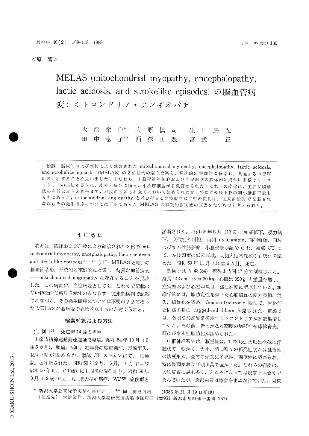
抄録 臨床的および剖検により確診されたmitochondrial myopathy, encephalopathy, lactic acidosis,and strokelike episodes (MELAS)の2剖検例の脳血管系を,系統的に電顕的に検索し,共通する血管病変の存在することを見い出した。すなわち,中膜平滑筋細胞および内皮細胞の胞体内に異常に多数のミトコンドリアの集積が見られ,変性・壊死に陥った平滑筋細胞が多数認められた。これらの変化は,主要な脳動脈の主幹部から末梢部まで,程度の差はあれ全てにおいて認められたが,特にクモ膜下腔の細小動脈で最も著明であった。mitochondrial angiopathyと呼び得るこの特徴的な血管の変化は,従来剖検例で記載されながらその発生機序については不明であったMELASの特徴的脳病変の原因をなすものと考えられた。
MELAS is a distinctive syndrome manifested by mitochodrial myopathy, encephalopathy, lactic acidosis, and recurrent stroke-like episodes such as seizures, alternating hemiparesis, hemianopsia, or cortical blindness. Pathologically the disorder is characterized by multiple, solitary or continuous foci of necrosis (infarct or softening), varying in size and stage, predominantly involving the bilateral cerebral cortices and to a lesser degree cerebral white matter, basal ganglia, brainstem and cereblum. The distribution of the lesions does not correspond to vascular territories, suggesting that they are not due to usual thrombotic or embolic process. The exact nature and pathoge-nesis of these lesions with characteristic distribu-tion pattern remain to be elucidated.
We studied systematically cerebral blood vessels from two autopsied patients with MELAS by electron microscopy. All the main cerebral arteries including anterior, middle and posterior cerebral, basilar and vertebral arteries were examined at their proximal portions at the cerebral base and at their peripheral portions at the cortical surface as well as within brain parenchyma.
We found marked accumulation of mitochondria in the cell bodies of smooth muscle cells and endothelial cells and numerous smooth muscle cells showing degeneration or necrosis, sporadically or in clusters in the tunics media. These abnorma-lities were most prominent in the walls of pial arterioles and small arteries up to 250μ in dia-meter, and less frequent and severe in the larger pial arteries and intracerebral arterioles and small arteries. These vascular changes are different from any of those described in various disorders known to involve the cerebral blood vessels and are thus characteristic to the cerebral blood vessels of MELAS.
We think that these peculiar vascular changes called mitochondrial angiopathy are caused by primary mitochodrial dysfunction in the vascular smooth muscle cells and endothelial cells them-selves, as is the same in the skeletal and cardiacmuscles in this disease, and that they constitute the pathogenic base of the brain lesions with unusual distribution pattern and nature in MELAS.

Copyright © 1988, Igaku-Shoin Ltd. All rights reserved.


