Japanese
English
- 有料閲覧
- Abstract 文献概要
- 1ページ目 Look Inside
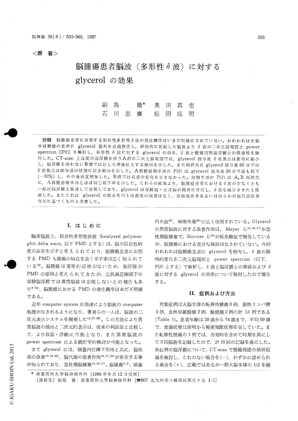
抄録 脳腫瘍患者に出現する限局性多形性δ波の発生機序はいまだ明確にされていない。われわれは大脳半球腫瘍の患者にglycerol製剤を点滴静注し,経時的に記録した脳波よりδ波の二次元脳電図とpowerspectrum ((Pδ))を解析し,多形性δ波に対するglycerolの効果,δ波と腫瘍周囲脳浮腫との関連性を検討した。CT-scan上高度の脳浮腫を伴うA群の二次元脳電図では,glycerol投与後δ波焦点は著明に縮小し,脳浮腫を伴わないB群ではむしろ増強拡大する傾向を示した。また両群共にglycerol投与後60分ではδ波焦点は投与前の状態に戻る傾向を示した。A群腫瘍側半球のP (δ)はglycerol投与後20分で最も低下(−30%)し,その後再度増加した。B群では有意の変化を示さなかった。対側半球のP (δ)はA,B両群共に,A群腫瘍側半球とほぼ同じ低下率を示した。これらの結果より,脳腫瘍患者におけるδ波の少なくとも一部は脳浮腫と関連して出現しており,glycerolは浮腫脳・正常脳の両者に作用し,δ波を減少させたと推測した。またこれはglycerolの脱水作用とは直接の関連はなく,脳血流改善あるいは何らかの脳代謝改善作用に基づくものと考察した。
The effect of intravenous infusion of 10% glycerol on polymorphic delta waves of EEG was investigated in 13 patients (15 studies) with brain tumors in cerebral hemisphere. The delta waves were analyzed by two-dimentional EEG topography and power spectrum of delta waves (P (δ)) over the tumor, adjacent, and distant regions in the same hemisphere to the tumor, and ipsi- and contralateral hemispheres to the tumor. The delta focus of EEG topography was well corresponded to the tumor and surrounding brain edema which visualized byx-ray CT-scan. In the patient with brain tumors with marked brain edema (group A, 11 studies), EEG topography showed marked reduction of delta focus within 20 minutes from the end of in-travenous infusion of glycerol, and returned to preinfusion distribution within 60 miutes. In pa-tients with absent to minimum brain edema (group B, 4 studies), delta focus showed the tendency to enlarge and returned to preinfusion state at 60 minutes after the end of glycerol infusion.
In patients of group A, P (δ) of ipsilateral hemi-sphere to tumor decreased at the end of glycerol infusion, and showing maximal reduction ratio (30%, p<0.01) at 20 minutes, re-increased there-after. In group B, P (δ) of ipsilateral hemisphere showed no definite changes after the glycerol infusion. P (δ) of contralateral hemispheres in patients of both group A and B, showed similar reduction ratio and time course to those of ipsi-lateral hemisphere of group A. In the ipsilateral hemisphere to tumor in group A, P (δ) over the tumor showed lowest reduction ratio, and highest at the distant region to the tumor by glycerol infusion. Eventhough statistically significant changes was absent in group B, P (δ) over the tumor showed tendency to increase and to decrease over the distant part to the tumor. When the patients were grouped according to presence or absence of midline shift on x-ray CT-scan, there was no sta-tistical difference of reduction ratio of P (δ) after glycerol infusion between two groups.
These results suggest that polymorphic delta waves appearing in patients with brain tumor of cerebral hemisphere originates, at least partially, from the functional disturbance of brain due to edema. It is also likely that delta waves in some part are related to mechanical effect of brain edema to midline structure, and disruption of corticosubcortical fiber connection induced by tumor itself.
Also, similar reduction ratios of P (δ) over the ipsilateral hemisphere to tumor in group A and contralateral hemispheres of group A and B, may suggest that the temporary decrease of delta waves by glycerol infusion is not the direct result of dehydrating effect of glycerol upon abnormally accumulated brain water, but of improvement and activation of cerebral blood flow or some me-tabolism of edematous and normal brain.

Copyright © 1987, Igaku-Shoin Ltd. All rights reserved.


