Japanese
English
- 有料閲覧
- Abstract 文献概要
- 1ページ目 Look Inside
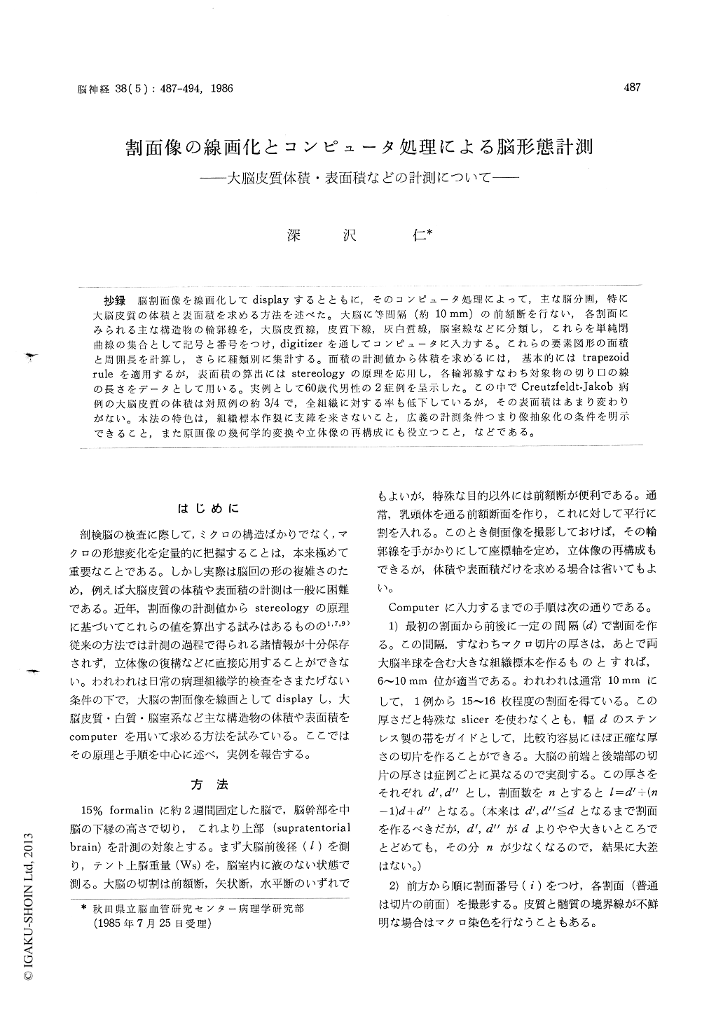
抄録 脳割面像を線画化してdisplayするとともに,そのコンピュータ処理によって,主な脳分画,特に大脳皮質の体積と表面積を求める方法を述べた。大脳に等間隔(約10mm)の前額断を行ない,各割面にみられる主な構造物の輸郭線を,大脳皮質線,皮質下線,灰白質線,脳室線などに分類し,これらを単純閉曲線の集合として記号と番号をつけ,digitizerを通してコンピュータに入力する。これらの要素図形の面積と周囲長を計算し,さらに種類別に集計する。面積の計測値から体積を求めるには,基本的にはtrapezoidruleを適用するが,表面積の算出にはstereologyの原理を応用し,各輪郭線すなわち対象物の切り口の線の長さをデータとして用いる。実例として60歳代男性の2症例を呈示した。この中でCreutzfeldt-Jakob病例の大脳皮質の体積は対照例の約3/4で,全組織に対する率も低下しているが,その表面積はあまり変わりがない。本法の特色は,組織標本作製に支障を来さないこと,広義の計測条件つまり像抽象化の条件を明示できること,また原画像の幾何学的変換や立休像の再構成にも役立つこと,などである。
A computer-aided morphometric method is des-cribed for evaluating the volumes and surface areas of brain structures from cut-surface images. This method consists of representing the topographical features of the cut surface as line drawings of closed curves and computing the magnitude of three-dimensional object from measurements made on two-dimensional sections.
After fixation in 15% formalin for at least two weeks, the supratentorial brain is cut coronally at intervals of d (approximately 10 mm). The thick-nesses of the extreme anterior and posterior slices are measured (d′, d″). All the cut surfaces are numbered (1≦i≦n), photographed, printed at a constant magnification of about 1.5, and the ana-tomical locations are specified on each print. The boundary lines of the main structures are classified into several categories : the cortical line, subcor-tical line, ventricular line, etc. Each of the classi-fied line is reduced to a set of simple closed cur-ves and entered into the computer in digitized form. The perimetric lengths as well as encircled areas of the constituent figures are computed and the classified totals of the boundary length andarea of brain structures are determined for indi-vidual cut surfaces. At the same time, the data are processed to be displayed on CRT as lines of different colors accordig to the classification.
The cerebral cortical volume is estimated from the series of its cut-surface using the trapezoidal rule. The cortical surface area (Sc) can be obtain-ed, on the assumption that the measurements are made with statistical uniformity, by the formula Sc=4/π{(n-1) d+d′ +d″} ΣLi/n, where Li is the length of cortical lines on each cut surface. This equation is derived from the basic equations of stereology.
In order to illustrate how the method is applied, two cases including a case of Creutzfeldt-Jakob disease are presented. It is suggested from the result that the surface area of cerebral cortex in Creutzfeldt-Jakob disease remains almost un-changed despite a marked decrease of the cortical volume.

Copyright © 1986, Igaku-Shoin Ltd. All rights reserved.


