Japanese
English
- 有料閲覧
PPV購入案内
1,320円 (1,200円+税10%) こちらは、973ページから980ページの文献です。購入には医書.jp本会員登録が必要です。
会員登録完了後に改めて上記「購入サイトへ」を選択してください。
または「購入サイトへ」選択後に「購入ログイン」、「新規会員登録」の順に進んでください。
会員登録について詳しくはをご確認ください。
- Abstract 文献概要
- 1ページ目 Look Inside
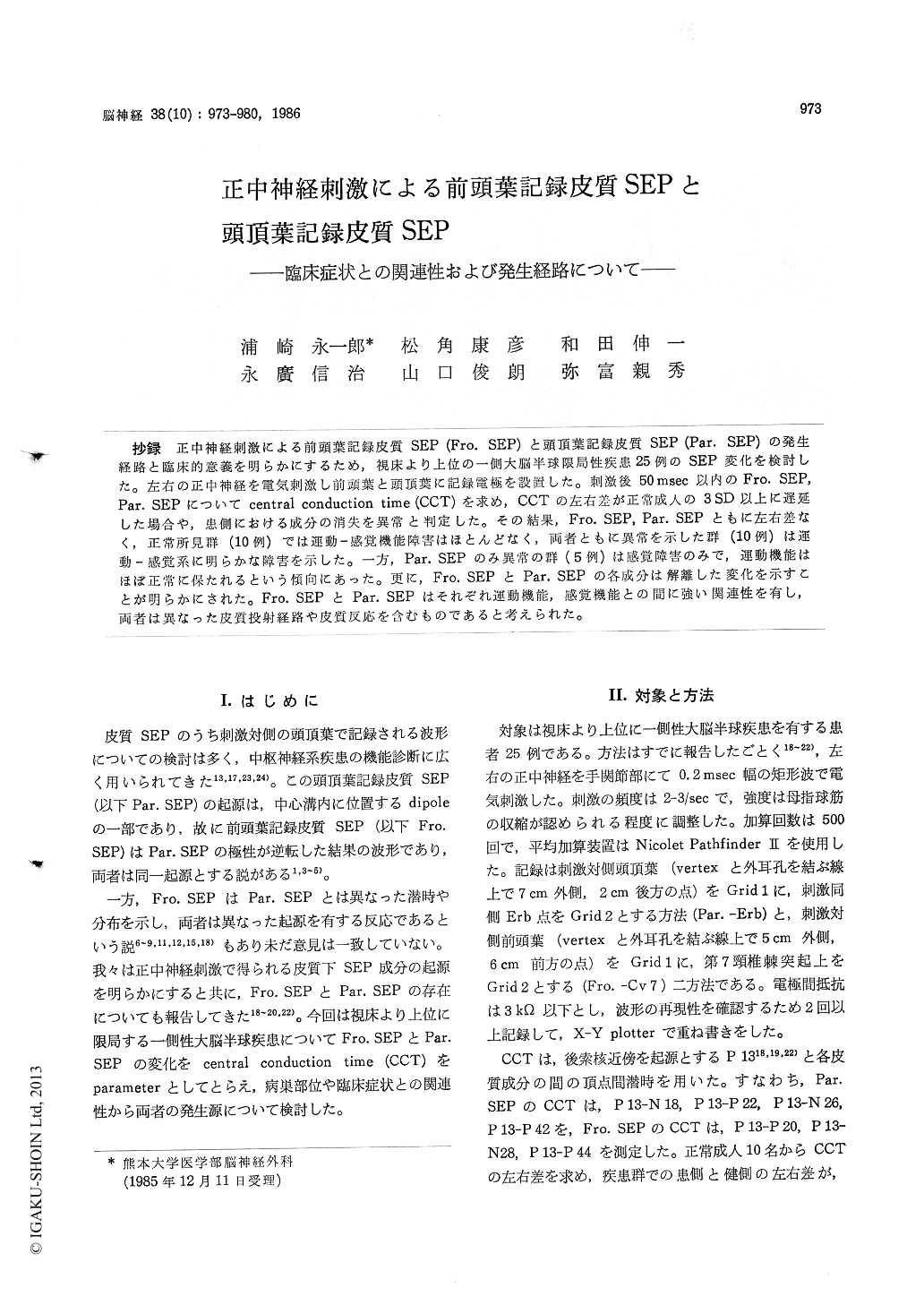
抄録 正中神経刺激による前頭葉記録皮質SEP (Fro. SEP)と頭頂葉記録皮質SEP (Par. SEP)の発生経路と臨床的意義を明らかにするため,視床より上位の一側大脳半球限局性疾患25例のSEP変化を検討した。左右の正中神経を電気刺激し前頭葉と頭頂葉に記録電極を設置した。刺激後50msec以内のFro. SEP,Par. SEPについてcentral conduction time (CCT)を求め,CCTの左右差が正常成人の3SD以上に遅延した場合や,患側における成分の消失を異常と判定した。その結果,Fro, SEP, Par. SEPともに左右差なく,正常所見群(10例)では運動—感覚機能障害はほとんどなく,両者ともに異常を示した群(10例)は運動—感覚系に明らかな障害を示した。一方,Par. SEPのみ異常の群(5例)は感覚障害のみで,運動機能はほぼ正常に保たれるという傾向にあった。更に,Fro. SEPとPar. SEPの各成分は解離した変化を示すことが明らかにされた。Fro. SEPとPar. SEPはそれぞれ運動機能,感覚機能との間に強い関連性を有し,両者は異なった皮質投射経路や皮質反応を含むものであると考えられた。
In order to know the chararteristics of frontal and parietal SEP components following median nerve stimulation, 25 patients with unilateral cerebral lesions above the thalamus were exami-ned, and their SSEPs were carefully compared with the clinical and radiological findings. In 10 normal subjects, there were three cortical compo-nents of the frontal SEPs (P 20-N 28-P 44) and four those components of the parietal SEPs (N 18-P 22-N 26-P 42). In patient's group, central conduction times (CCTs) between component P 13 and each cortical component were measured and the latency differences between normal side and affected side were calculated. When the latency differences increased over 3 S. D. from the mean of the control values or the some cortical compo-nents disappeared, they were regarded as abnor-mal. According to the combination of the abnor-malities in frontal and parietal SEPs, three groups were classified as follows : group 1 ; frontal and parietal SEPs were normal (n =- 10), group 2 ; frontal and parietal SEPs were both affected (n=10), group 3; parietal SEPs were affected but frontal components were preserved in normal range (n= 5 ). CT scan showed that the region from internal capsule to cortex around the central sulcus remained intact in the patients of group 1, while this region was involved in various degrees in all cases of the group 2. In patients of group 3, frontal or parietal regions were variously affected. Both the motor and sensory functions were mainly intact in group 1, and disturbed in group 2. In group 3, only sensory disturbances were revealed. Furthermore, the dissociate changes between frontal SEPs and parietal SEPs were apparent in the some cases of group 2 and 3. All of these findings suggested that the frontal P 30-N 28-P 44 were independent from the parietal N 18-P 22-N 26-P 42. This study also indicated good correlations between motor function and frontal SEPs, and sensory function and parietal SEPs, respectively.
Copyright © 1986, Igaku-Shoin Ltd. All rights reserved.