Japanese
English
- 有料閲覧
- Abstract 文献概要
- 1ページ目 Look Inside
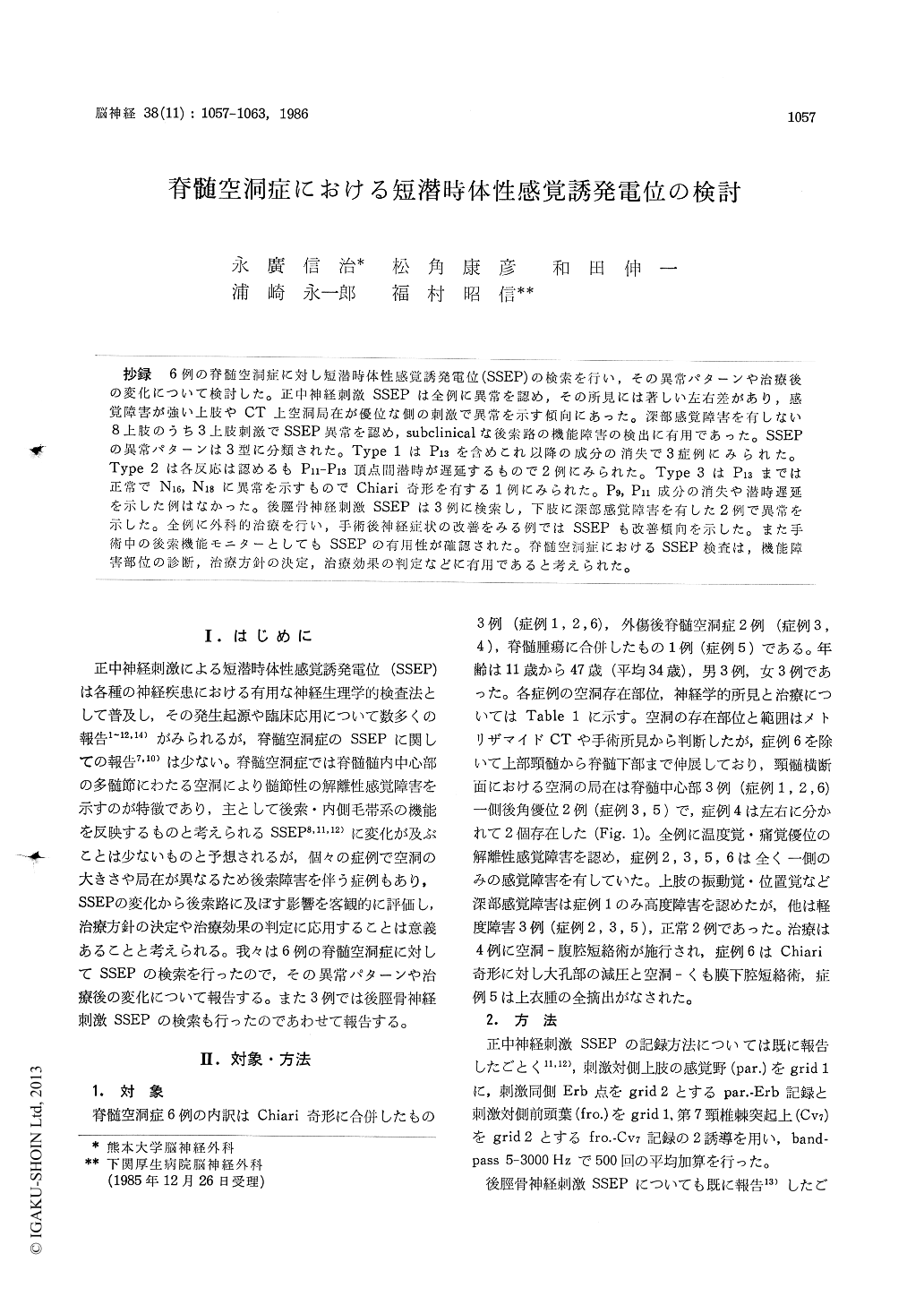
抄録 6例の脊髄空洞症に対し短潜時体性感覚誘発電位(SSEP)の検索を行い,その異常パターンや治療後の変化について検討した。正中神経刺激SSEPは全例に異常を認め,その所見には著しい左右差があり,感覚障害が強い上肢やCT上空洞局在が優位な側の刺激で異常を示す傾向にあった。深部感覚障害を有しない8上肢のうち3上肢刺激でSSEP異常を認め,subclinicalな後索路の機能障害の検出に有用であった。SSEPの異常パターンは3型に分類された。Type 1はP13を含めこれ以降の成分の消失で3症例にみられた。Type 2は各反応は認めるもP11—P13頂点間潜時が遅延するもので2例にみられた。Type 3はP13までは正常でN16, N18に異常を示すものでChiari奇形を有する1例にみられた。P9, P11成分の消失や潜時遅延を示した例はなかった。後脛骨神経刺激SSEPは3例に検索し,下肢に深部感覚障害を有した2例で異常を示した。全例に外科的治療を行い,手術後神経症状の改善をみる例ではSSEPも改善傾向を示した。また手術中の後索機能モニターとしてもSSEPの有用性が確認された。脊髄空洞症におけるSSEP検査は,機能障害部位の診断,治療方針の決定,治療効果の判定などに有用であると考えられた。
Short latency somatosensory evoked potentials (SSEPs) following median nerve and posteriortibial nerve stimulation were studied in six pa-tients with syringomyelia. Three patients had Chiari malformations, two patients experienced fracture of the spine and one patient had a cauda equina ependymoma.
SSEPs following median nerve stimulation were abnormal in all patients, of which five patients showed abnormal SSEPs only in the unilateral stimulation on the side of sensory deficits. SSEPs obtained from three out of eight upper extremities which showed no disturbance of deep sensation, were abnormal, so SSEPs were able to detect subcli-nical abnormality indicating dorsal column dysfunc-tion. Abnormal patterns of SSEPs were classified in three types as followes; Type 1: disappearance of P13, N16 and N18 (3 cases), Type 2: the prolonged interpeak latency P11-P13 (2 cases), and Type 3: abnormal N16 and N18 with preserving P13 (1 case with Chiari malformation). P9 and P11 were pre-sent without prolonged latencies in all cases.
SSEPs following posterior tibial nerve stimula-tion were abnormal in two of the three tested patients. Those two patients had disturbance of deep sensations in the lower extremities.
All patients underwent surgical treatment, syringo-peritoneal shunt in four patients, foramen magnum decomopression with syringo-subarach-noid shunt in one patient, and total removal of an ependymoma of the cauda equina with syringo-stomy in one patient. Postoperative neurological improvement were found in three patients, of which two cases also showed improvement in SSEPs. On the contrary SSEPs were unchanged in two patients with posttraumatic syringomyelia, whose postoperative neurological condition was also unchanged. SSEPs were directly recorded from the cuneate nucleus for the intraoperative monitoring during syringostomy in one patient.
Analysis of SSEPs in patients with syringomylia is useful for evaluating the function of the dorsal column to the medial lemniscus before, during and after surgery.

Copyright © 1986, Igaku-Shoin Ltd. All rights reserved.


