Japanese
English
- 有料閲覧
- Abstract 文献概要
- 1ページ目 Look Inside
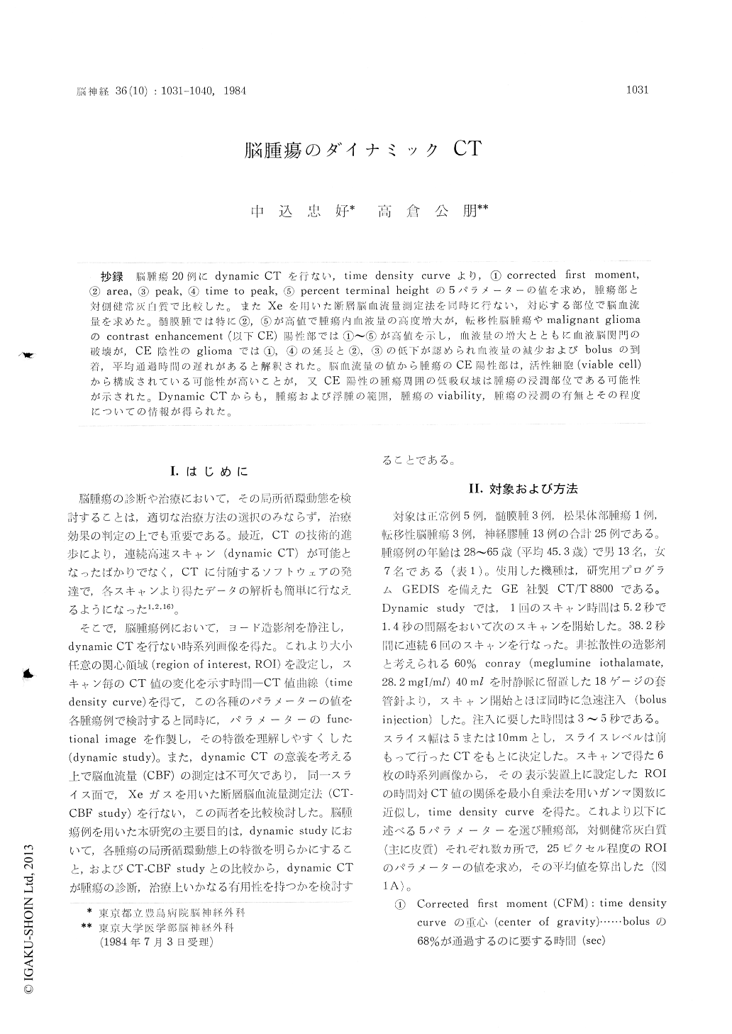
抄録 脳腫瘍20例にdynamic CTを行ない,time density curveより,①corrected first moment,②area,③peak,④time to peak,⑤percent terminal heightの5パラメーターの値を求め,腫瘍部と対側健常灰自質で比較した。またXeを用いた断層脳血流量測定法を同時に行ない,対応する部位で脳血流量を求めた。髄膜腫では特に②,⑤が高値で腫瘍内血液量の高度増大が,転移性脳腫瘍やmalignant gliomaのcontrast enhancement (以下CE)陽性部では①〜③が高値を示し,血液量の増大とともに血腋脳関門の破壊が,CE陰性のglioInaでは①,④の延長と②,③の低下が認められ血液量の減少およびbolusの到着,平均通過時間の遅れがあると解釈された。脳血流量の値から腫瘍のCE陽性部は,活性細胞(viable cell)から構成されている可能性が高いことが,又CE陽性の腫瘍周囲の低吸収域は腫瘍の浸潤部位である可能性が示された。Dynamic CTからも,腫瘍および浮腫の範囲,腫瘍のviability,腫瘍の浸潤の有無とその程度についての情報が得られた。
Dynamic computed tomography (CT) has been widely used because of its simplicity, but regional cerebral perfusion of brain tumors has not critically been evaluated. This study was conducted to evaluated parameters obtained from the dynamic perfusion study in brain tumors and to clarify the usefulness of the dynamic CT comparing with Xe-enhanced CT.
Dynamic CT was performed on 20 patients with brain tumor (three meningiomas, one pineal tumor, three metastatic brain tumors, thirteen gliomas). Dynamic CT consisted of performing six rapid sequential scans after a bolus intravenous injection of 40 ml of iodinated contrast medium. In this study, five parameters (corrected firstmoment, area, peak, time to peak, percent terminal height) were obtained from computer analysed curve fit on time density curve profile of serial scanning. In Xe-enhanced CT, serial CT was taken every three or five minutes during inhalation of 40 to 50 percent stable xenon. Flow rate con-stant (K), partition coefficient (λ) and cerebral blood flow (CBF) for each pixel were calculated from ΔHU (Hounsfield unit) and end-tidal air curve and displayed on CRT as images.
In patients with meningioma, values of area and percent terminal height of the tumor were much higher than those of contralateral gray matter, and these findings show excessive increase of the intratumoral blood volume and extreme extravasation of iodinated contrast medium respectively.
In patients with metastatic brain tumor or malignant glioma, which were enhanced by con-trast medium, the values of all parameters of the tumors were higher than contralateral gray matter. Increase of intratumoral blood volumeand destruction of blood brain barrier are sugge-sted in these tumors.
In the remaining patients with glioma, which was not enhanced, the values of corrected first moment and time to peak of the tumors were higher but area and peak were lower than contra-lateral gray matter. These findings show decrease of intratumoral blood volume and delay of mean transit time and arrival time of the bolus.
Compared with CBF measured by Xe-enhanced CT, it was suggested that high density area of metastatic brain tumor or malignant glioma con-sisted of viable tumor cells, and in patients with glioma, low density area adjacent to the tumor with contrast enhancement was the invasive site of the tumor.
Using dynamic CT, we were also able to distinguish the tissue contains viable tumor cells from the other part of brain by the differences in the parameter value.

Copyright © 1984, Igaku-Shoin Ltd. All rights reserved.


