Japanese
English
- 有料閲覧
PPV購入案内
1,320円 (1,200円+税10%) こちらは、583ページから594ページの文献です。購入には医書.jp本会員登録が必要です。
会員登録完了後に改めて上記「購入サイトへ」を選択してください。
または「購入サイトへ」選択後に「購入ログイン」、「新規会員登録」の順に進んでください。
会員登録について詳しくはをご確認ください。
- Abstract 文献概要
- 1ページ目 Look Inside
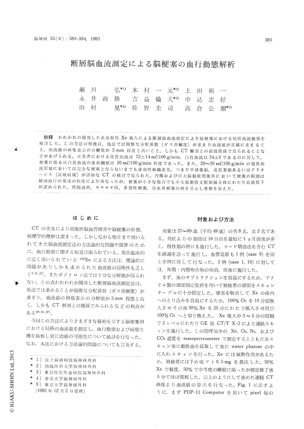
抄録 われわれの開発した非放射性Xe吸入による断層脳血流測定により脳梗塞における局所血流動態を検討した。この方法の特徴は,他法では困難な分配係数(ガス溶解度)が求まり血流値が正確に求まること,血流値の画像表示の分解能が3mm程度と高いこと,しかもCT解剖との直接関係で見られることなどがあげられる。正常者における皮質血流は72±14ml/100g/min,白質血流は24±5であるのに対して,梗塞に陥る灰白質血流の虚血閾値は20ml/lOOg/min程度であった。また,20〜30ml/100g/minの境界血流領域においては完全な梗塞とならないまでも虚血性組織変化,つまり半球萎縮,皮質萎縮あるいはグリオーシス(高吸収域)が詳細なCTの検討で見られた。内頸および中大脳動脈閉塞例において梗塞の範囲は側副血行の発達の程度により異なったが,梗塞が小さな場合でも中大脳動脈支配領域全体にわたり血流低下が認められた。再開通例,モヤモヤ病,多発性梗塞,分水界梗塞の例を呈示し考察を加えた。
Cerebral perfusion was examined in various types of occlusive disease by computed tomogra-phic CBF method. The method utilized has several advantages over conventional studies using iso-tope, providing high resolution images in a direct relation to CT anatomy. Ten representative cases were presented from 25 consective cases of occlu-sive disease studied by this method. The method included inhalation of 40 to 60% xenon with serial CT scanning for 25 min. K (build-up rate), λ (partition coefficient) and CBF values were cal-culated from JHU for each pixel and JXe in ex-pired air, based on Fick's principle, and dis-played on CRT as K-, A- and CBF-map separa-tely. CBF for gray matter of normal control was 82±11 ml/100 gm/min and that for white matter was 24±5 ml/100 gm/min.
The ischemic threshold for gray matter appear-ed to be approximately 20 ml/100 gm/min, as blood flow in focus of complete infarction was below this level. Blood flow between 20-30 ml/ 100 gm/min caused some change on CT, such as localized atrophy, cortical thinning, loss of distinc-tion between gray and white matter and decreas-ed or increased density, which were considered to be compatible with pathological changes of laminar necrosis or gliosis with neuronal loss.
In a case with occlusion of middle cerebral artery with subsequent recanalization, causing hemorrhagic infarct, hyperemia was observed in the infarcted cortex that was enhanced by iodine. Periventricular lucency observed in two cases, where blood flow was decreased below threshold, could be classified as "watershed infarction" main-ly involving white matter. In moyamoya disease, blood flow in the anterior circulation was decre-ased near ischemic level, whereas that in basal ganglia and territory of posterior cerebral artery was fairly preserved, which was compatible with general angiographic finding of this disease.
Copyright © 1983, Igaku-Shoin Ltd. All rights reserved.