Japanese
English
- 有料閲覧
- Abstract 文献概要
- 1ページ目 Look Inside
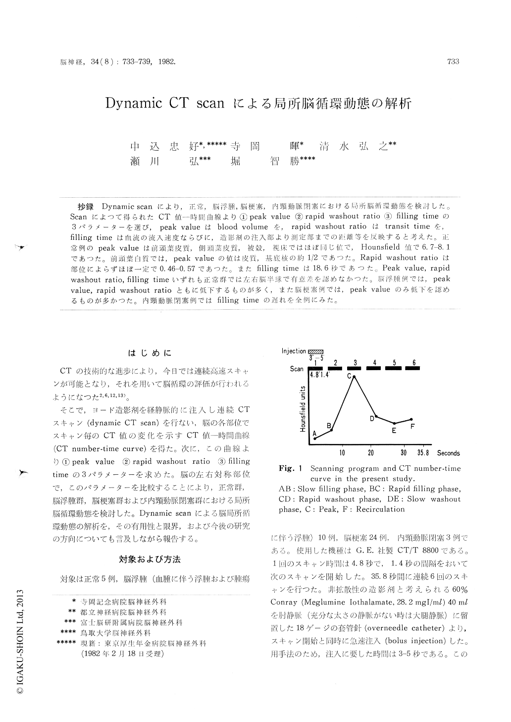
抄録 Dynamic scanにより,正常,脳浮腫,脳梗塞,内頸動脈閉塞における局所脳循環動態を検討した。Scanによつて得られたCT値—時間曲線より①peak value②rapid washout ratio③filling timeの3パラメーターを選び,peak valueはblood volumeを,rapid washout ratioはtransit timeを,filling timeは血流の流入速度ならびに,造影剤の注入部より測定部までの距離等を反映すると考えた。正常例のpeak valueは前頭葉皮質,側頭葉皮質,被殻,視床ではほぼ同じ値で,Hounsfield値で6.7-8.1であつた。前頭葉白質では,peak valueの値は皮質,基底核の約1/2であつた。Rapid washout ratioは部位によらずほぼ一定で0.46—O.57であつた。またfilling timeは18.6秒であつた。Peak value,rapidwashout ratio,filling timeいずれも正常群では左右脳半球で有意差を認めなかつた。脳浮腫例では,peak value,rapid washout ratioともに低下するものが多く,また脳梗塞例では,peak valueのみ低下を得認めるものが多かつた。内頸動脈閉塞例ではfilling timeの遅れを全例にみた。
Regional brain circulation was evaluated by dynamic computed tomography with iodine enhan-cement. The CT number (Hounsfield unit)-time curve provided us three parameters, namely, peak value, rapid washout ratio and filling time. It was indicated that these three parameters were related to different components. Peak value wasrelated to blood volume, rapid washout ratio to transit time and filling time to filling velosity and/or distance from the point of injection to the site of measurement. In 10 patients with brain edema, both peak value and rapid washout ratio were decreased, comparing to these of 5 normal subjects. In 24 patients with cerebral in-farction, decreased peak value was a main finding. In 3 patients with occlusion of internal carotid artery, delayed filling time was demonstrated in every patients. Clinical application of dynamic computed tomography was discussed.

Copyright © 1982, Igaku-Shoin Ltd. All rights reserved.


