Japanese
English
- 有料閲覧
- Abstract 文献概要
- 1ページ目 Look Inside
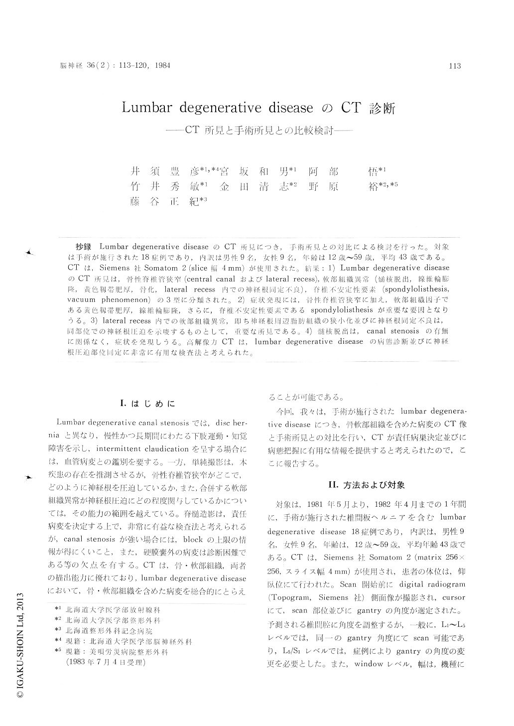
抄録 Lumbar degenerative diseaseのCT所見につき,手術所見との対比による検討を行った。対象は手術が施行された18症例であり,内訳は男性9名,女性9名,年齢は12歳〜59歳,平均43歳である。CTは,Siemens社 Somatom 2 (slice幅4mm)が使用された。結果:1) Lumbar degenerative diseaseのCT所見は,骨性脊椎管狭窄(central canalおよびlateral recess),軟部組織異常(髄核脱出,線維輪膨隆,黄色靱帯肥厚,骨化, lateral recess 内での神経根同定不良),脊椎不定性要素(spondylolisthesis,vacuum phenomenon)の3型に分類された。2)症状発現には,骨性脊椎管狭窄に加え,軟部組織因子である黄色靱帯肥厚,線維輪膨隆,さらに,脊椎不安定性要素である spondylolisthesisが重要な要因となりうる。3) lateral recess内での軟吹部組織異常,即ち神経根周辺脂組織の狭小化並びに神経根同定不良は,同部位での神経根圧迫を示唆するものとして,重要な所見である。4)髄核脱出は,canal stenosisの 有無に関係なく,症状を発現しうる。高解像力CTは,lumbar degenerative diseaseの病態診断並びに神経根圧迫部位同定に非常に有用な検査去と考えられた。
We reported the 18 patients which underwent surgical exploration and reviewed these CT find-ings.
Method
All CT scans were obtained on Somatom II, high resolution CT scanner, with the patient in the supine position. A lateral localizer image (To-pogram) was used to select the appropriate inter-vertebral disk space. The slice thickness was 4 mm.
Results
1) CT findings in lumbar degenerative diseases include bony canal stenosis (central canal stenosis,narrowed lateral recess), soft tissue abnormalities (herniated nucleus pulposus, bulging annulus, hypertrophy and/or ossification of ligamentum fla-vum, no delineation of nerve root in lateral recess), and spinal instability (spondylolisthesis, vacuum phenomenon).
2) The above three factors contribute to nar-rowing of spinal canal.
3) No delineation of nerve root or soft tissue replacement of epidural fat in lateral recess sug-gests that the nerve root may be compressed by some factors.
4) Ilerniated nucleus pulposus may cause nerve root compression with or without canal stenosis.
Conclusion
This study revealed that the CT findings corre-lated closely with the surgical findings and the site of nerve root compression could be determi-ned.

Copyright © 1984, Igaku-Shoin Ltd. All rights reserved.


