Japanese
English
- 有料閲覧
- Abstract 文献概要
- 1ページ目 Look Inside
I.はじめに
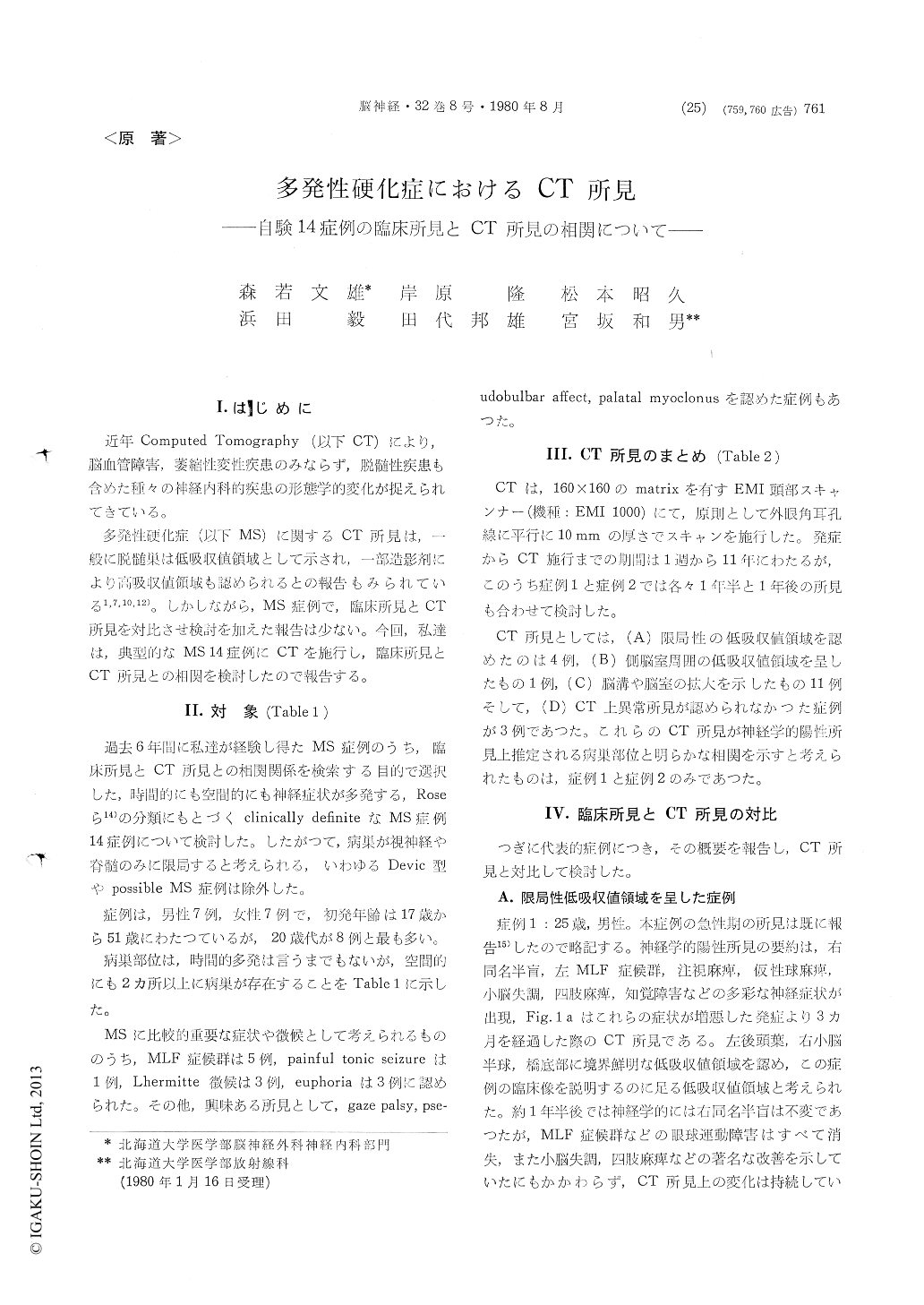
近年Computed Tomography (以下CT)により,脳血管障害,萎縮性変性疾患のみならず,脱髄性疾患も含めた種々の神経内科的疾患の形態学的変化が捉えられてきている。
多発性硬化症(以下MS)に関するCT所見は,一般に脱髄巣は低吸収値領城として示され,一部造影剤により高吸収値領域も認められるとの報告もみられている1,7,10,12)。しかしながら,MS症例で,臨床所見とCT所見を対比させ検討を加えた報告は少ない。今回,私達は,典型的なMS 14症例にCTを施行し,臨床所見とCT所見との相関を検討したので報告する。
Recent introduction of Computed Tomography (CT) in clinical neurology made it possible to visualize the brain lesion without any invasive procedures.
In multiple sclerosis (MS), the demyelinating foci were reported to be observed as low density areas on CT, but occasionally contrast enhanced high density areas were reported also. So far as we know, only a few reports which analysed inter-relationship between clinical signs and CT findings were published.
In this report we tried to examine the correlation of clinical findings with CT in MS. All scans were performed using an EMI head scanner (EMI 1000) with a 160×160 matrix. Contrast material was administered as an intravenous bolus of 60% meg-lumine iothalamate.
In clinically definite 14 MS patients, CT showed localized, circumcribed low density areas in 4 patients, periventricular low density in 1 patient, widening of cortical sulci with ventricular dila-tation in 11 patients and no abnormalities in 3 patients.
The widening of cortical sulci with ventricular dilatation were noted to be particularly common findings.
The periventricular low density was not so frequently seen as we expected.
Localized, circumscribed low density areas on CT were well correlated with the neurological findings in 2 patients. In these cases the abnormalities on CT persisted in spite of neurological improvement.
As a conclusion, we think CT might be useful as a diagnostic evaluation of MS.

Copyright © 1980, Igaku-Shoin Ltd. All rights reserved.


