Japanese
English
- 有料閲覧
- Abstract 文献概要
- 1ページ目 Look Inside
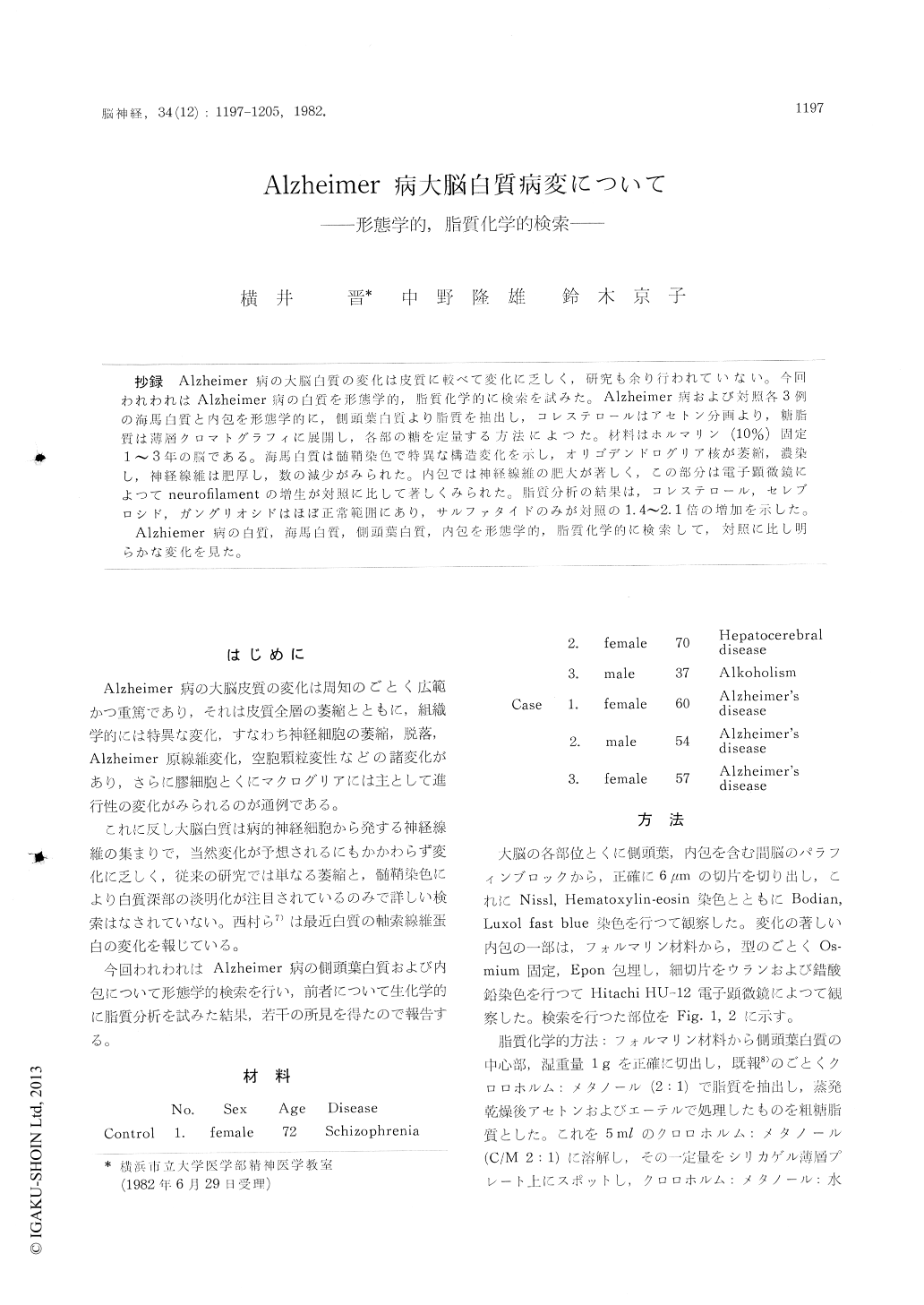
抄録 Alzheimer病の大脳白質の変化は皮質に較べて変化に乏しく,研究も余り行われていない。今回われわれはAlzheimer病の白質を形態学的,脂質化学的に検索を試みた。Alzheimer病および対照各3例の海馬白質と内包を形態学的に,側頭葉白質より脂質を抽出し,コレステロールはアセトン分画より,糖脂質は薄層クロマトグラフィに展開し,各部の糖を定量する方法によつた。材料はホルマリン(10%)固定1〜3年の脳である。海馬白質は髄鞘染色で特異な構造変化を示し,オリゴデンドログリア核が萎縮,濃染し,神経線維は肥厚し,数の減少がみられた。内包では神経線維の肥大が著しく,この部分は電子顕微鏡によつてneurofilamentの増生が対照に比して著しくみられた。脂質分折の結果は,コレステロール,セレブロシド,ガングリオシドはほぼ正常範囲にあり,サルファタイドのみが対照の1.4〜2.1倍の増加を示した。
Alzhiemer病の白質,海馬白質,側頭葉白質,内包を形態学的,脂質化学的に検索して,対照に比し明らかな変化を見た。
The cerebral white matter of Alzheimer's disease were investigated neuropathologically and lipid-chemically. The materials were composed of the brains of 3 cases of Alzheimer's disease and of 3 cases of control of various diseases, which were stored in 10% formalin for 1-3 years.
In this study, the white matter of the ammons horn and internal capsule were chosen for the morphological examinations and the white matter of the temporal lobe for lipid-chemical analysis (Fig. 1, 2), because the temporal lobe including ammons horn is a predilection site of pathological senile processes and the internal capsule is an assemblate of descending tracts.
In myelin preparations, the white matter of the ammons horn showed characteristic changes of myelin sheath associated with an increase of shrinked nuclei of oligodendro glia cells. The thickening and rarefication of nerve fibers in the white matter of ammons horn were observed by Bodian staining (Fig. 3, 4, 5, 6).
Severe thickening and deformation were demon-strated in nerve fibers of the internal capsule of the cases of Alzheimer's disease, although thesame changes were slightly observed in control cases (Fig. 7, 8). Marked proliferation of neurofila-ments was noticed in nerve fibers of the internal capsule under electronmicroscopy but there were no abnormal increase of mitochondria, deposition of dense body and conglomerate of neurofilaments (Fig. 9, 10). This finding was in agreement with the findings of Nishimura et al which proved the increase of neurofilament-like protein in the white matter of Alzheimer's disease biochemically.
Lipid analysis : Cholesterol and glycolipids were extracted from the white matter of temporal lobe following the method previously reported. Crude glycolipids were developed on a slica gel thin layer plate by solvent system of chloroform : methanol: water (65: 25 : 4) and the spots cor-responding to cerebroside, sulfatide and ganglio-side were scraped off and the volume of sugar in them were determined by anthrone reagent. Total cholesterol was estimated by Zak-Henly method. The results of the determinations ofcholesterol and glycolipids were listed in Table 1. Comparing with control cases, the values of cholesterol, cerebroside and ganglioside were within normal range, however sulfatide showed a marked increase of 1. 4-2. 1 times as much as that of control. The ratio of cerebroside to sul-fatide of 3 cases of Alzheimer's disease was 1. 5- 2. 1, while that of control was 3. 2. Cherayil and Cyrus reported the quantitative estimation of glycolipids in the brain of Alzheimer's disease. In one of their 3 cases, sulfatide in the midfrontal white matter showed an elevation and the ratio of cerebroside to sulfatide was 2. 5.
The senile plaque of Alzheimer's disease shows histochemical character of so-called amyloid, which contains much sulfated polyglucose. Our findings of increase of sulfatide in the temporal white matter suggests that there may exist some abnor-mal metabolism of sulfate in the brain of this disease.

Copyright © 1982, Igaku-Shoin Ltd. All rights reserved.


