Japanese
English
- 有料閲覧
- Abstract 文献概要
- 1ページ目 Look Inside
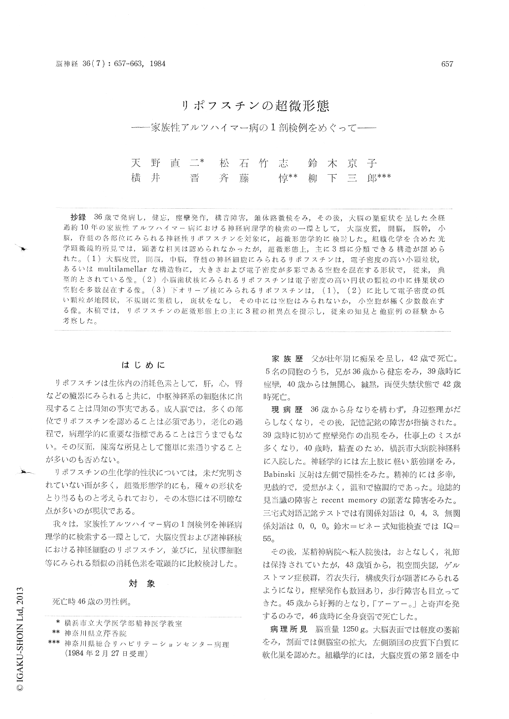
抄録 36歳で発病し,健忘,痙攣発作,構音障害,錐体路徴候をみ,その後,大脳の巣症状を呈した全経過約10年の家族性アルツハイマー病における神経病理学的検索の一環として,大脳皮質,間脳,脳幹,小脳,脊髄の各部位にみられる神経性リポフスチンを対象に,超微形態学的に検討した。組織化学を含めた光学顕微鏡的所見では,顕著な相異は認められなかったが,超微形態上,主に3群に分類できる構造が認められた。(1)大脳皮質,間脳,中脳,脊髄の神経細胞にみられるリポフスチンは,電子密度の高い小顆粒状,あるいはmultilamellarな構造物に,大きさおよび電子密度が多彩である空胞を混在する形状で,従来,典型的とされている像。(2)小脳歯状核にみられるリポフスチンは電子密度の高い円状の顆粒の中に蜂巣状の空胞を多数混在する像。(3)下オリーブ核にみられるリポフスチンは,(1),(2)に比して電子密度の低い顆粒が地図状,不規則に集積し,斑状をなし,その中には空胞はみられないか,小空胞が極く少数散在する像。本稿では,リポフスチンの超微形態上の主に3種の相異点を提示し,従来の知見と他症例の経験から考察した。
This work is to study the ultrastructure of the neuronal lipofuscin that occurred in the brain and the spinal cord of an autopsy case of familial Alzheimer's disease and to compare with those in several other diseases.
The patient was a 46-year-old male, whose fa-ther and elder brother were diagnosed as Alzhei-mer's disease and died at the age of 42, respec-tively. He became afflicted with forgetfulness and disorientation at the age of 36. He developed a grand mal seizure at the age of 39 and thereafter, his clinical course was characterized by pyramidal signs, dysarthria and the symptoms of Gerstmann's symdrome, visuo-spatial agnosia, apraxia for dres-sing and constructive apraxia. He became bedrid-den at 45 years old and died of general prostra-tion.
The brain weighed 1, 250g, and the cerebral cortex showed mild atrophy. The neuronal loss, senile plaques and Alzheimer's neurofibrillary tangles were found throughout the cerebral cortex. The senile plaques were also found in the basal ganglia, the cerebellar medulla and cortex. There was severe amyloid angiopathy in the occipital and cerebellar cortices.
The specimens for electron microscopy weretaken from the cerebral cortex, the basal ganglia, the thalamus, the midbrain, the medulla oblongata, the cerebellum and the spinal cord. The ultra-structural study revealed three different types of the neuronal lipofuscin, though different stain-ability between these lipofuscin granules could not be manifested by several histochemical me-thods. Their morphological differences seemed to be based on the sites of the central nervous sys-tem. Three types are clarified as follows:
1) In the cerebral cortex, the striatum, the lat-eral geniculate body, the thalamus and the anterior horn, typical lipofuscin granules were observed. They were made up of irregularly shaped, gran-ular components accompanied with vacuoles which greatly varied in size and electron density. The granular components consisted of multilamellar or/and dotted structures.
2) In the dentate nucleus of the cerebellum, the lipofuscin consisted of round, electron-dense gra-nules including many honeycomb-like vacuoles.
3) In the inferior olivary nucleus, the lipofuscin was composed of irregularly shaped, mottled con-figurations which were rather amorphous and low in electron density. Vacuoles were rarely seen.
The ultrastructural differences of the neuronal lipofuscin in the different neurons were demon-strated in this case and their implications were discussed refering to the literature.

Copyright © 1984, Igaku-Shoin Ltd. All rights reserved.


