Japanese
English
- 有料閲覧
- Abstract 文献概要
- 1ページ目 Look Inside
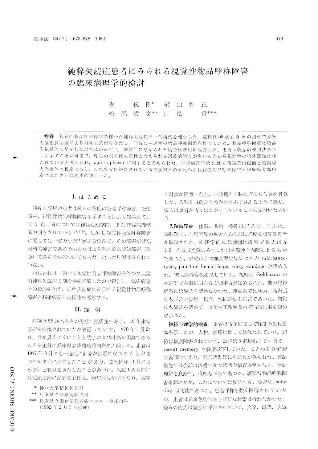
抄録 視覚性物品呼称障害を伴った純粋失読症の一剖検例を報告した。症例は58歳右ききの男性で左後大脳動脈閉塞により純粋失読症をきたし,同時に一過性の物品呼称困難を伴つていた。物品呼称困難は物品を視覚的に呈示した場合にのみ生じ,触覚的に与えられた場合は著明に改善した。患者は物品の使用法を正しく示すことが可能で,呼称の誤りは失語性と考えられる語義的誤りが多いことから視覚性の物体認知は保たれていると考えられ,optic aphasiaに属すると考えられた。神経病理学的には左後頭葉内側面と脳梁膨大部全体の梗塞であり,これまでに報告されている剖検例との対比から視覚性物品呼称障害と脳梁膨大部病変の大きさとの関連に注目した。
It has long been known that some of pure alexic have trouble with visual object naming. However, the question whether the defect represents anomic errors or more basic perceptive errors (agnosia) remains unsettled and the responsible lesions for it have never been provided.
We report here a typical case of alexia without agraphia and with transient anomia for visually presented objects. Clinicoanatomical correlation led us to a speculation that vision specific anomia without agnosia can be correlated with spleniallesion.
A 58 year-old right-handed man, a drugstore keeper, had been known to be diabetic for 10 years. On December 1977, he suffered from severe head-ache on the left parietal region and also felt forgetfulness and difficulty of reading and calcula-tion. He was admitted on January 24th, 1978. On examination, there was dense right-sided hemi-anopia of the left eye. Visual acuity was 10/10 O. S. and n. d. O. D. because of traumatic cataract. The rests of the cranial nerves, motor systems, sensory modalities and reflexes were normal. Con-versational speech was almost normal. Reading ability was completely impaired. There was no difference among letters, words and sentenses, or among Kana, Kanji and digits. Writing was sig-nificantly superior to reading. Kinesthetic reading was considerably better. Copying of written ma-terials was also impaired. Naming of colors was quite inappropriate but full investigation was not performed because he happened to be color blind.
There was a difficulty in naming familiar objects. Twenty familiar objects were presented to the patient. He was requested to name them visually. When he failed to name an object, it was handed to him. He was able to name nine objects visually without or with phonetic cuing. Most of the naming errors were semantic, for example, he misnamed a pencil as a fountain pen and a crip as a pin. He was explained correctly how to use the misnamed object and made appropriate definition (periphrase). For instance, a pocket light was replied as" doctors use it to observe patients' eyes". He could also gesticulate how to use the misnamed object. Once objects were handed to the patient,he could name all except two.
Six months later, object naming showed signi-ficant improvement, but alexic state not at all. The patient died a year later because of pontine hemor-rhage, and autopsy was obtained. The organized thrombi occluded the left posterior cerebral artery. Beside a gross hemorrhage in the pons, there was an old infarction in the mesial aspect of the left occipito-temporal lobe, i.e., the whole of the lingual and fusiform gyrus, inferior half of the cuneus, posterior part of the hippocampal and the parahip-pocampal gyrus, crus of the fornix and the whole of the forceps major and the splenium of the corpus callosum.
In the present case of typical alexia without agraphia, the anomia is strictly visual and improved through tactile presentation. The fact that he could either demonstrate or describe the nature of an object proved preserved visual recognition. Thus the defect belongs to vision specific anomia or optic aphasia rather than visual agnosia. The neuro-pathology of the present case is typical for that of classical pure alexia. We reviewed the liter-ature of pure alexia and divided the cases into anomic cases and non-anomic cases. The com-parison of neuropathology of these two groups revealed that the only difference is the extent of the splenium involvement. in the anomic group, the splenium involvement is total while in the non-anomic group, it remains incomplete. Thus it could be speculated that portion of the intact sple-nium secures interhemispheric visual-verbal associa-tion and that the total splenium destruction is responsible for vision specific anomia in pure alexia.

Copyright © 1982, Igaku-Shoin Ltd. All rights reserved.


