Japanese
English
- 有料閲覧
- Abstract 文献概要
- 1ページ目 Look Inside
I.はじめに
虚血性脳血管障害例において,その臨床症状の帰結を頭蓋内血行動態におけるsteal mechanism18,19)に求めることは厳密にはしばしば困難なことが多い。しかし内頸動脈(IC)系の閉塞性障害に伴つて椎骨脳底動脈(V-B)系の一過性脳虚血発作(TIA),または椎骨脳底動脈循環不全(VBI)に相当する症状が認められることは決してまれではない2,3,7,10)。著者らは神経放射線学的所見,臨床経過よりV-B系からIC系へintracerebral stealが生じたと考えられる一側総頸動脈狭窄例を経験した。本例は,比較的まれとされる後頭動脈—椎骨動脈(OA-VA)吻合が症状発現に関与しており,総頸動脈内膜剥離術(CEA)前後の血行動態の変化から本吻合の側副路としての意義と,intracerebral steal現象について文献的考察を加えて報告したい。
A case of vertebro-basilar insufficiency, with symptoms released by common carotid endarterec-tomy, is reported. The etiology of this case, proba-bly due to intracerebral steal mechanisms, may be explained based on historical, CT and angiographic findings of the pre- and postoperative states as available.
This 54-year-old man, having a history of mild Diabetes Mellitus and artial fibrillation, was ad-mitted to our hospital following repeated ischemic attacks during the previous two years.
These ischemic episodes included fainting sensa-tion, vertigo and numbness of the right hand. At the same time, headaches were recognized during these episodes, and during the last attack, truncal ataxia was detected in addition to the other symptoms. The combination of these symptoms might coincide with a diagnosis of transient is-chemic attacks in the vertebro-basilar system, or vertebro-basilar insufficiency.
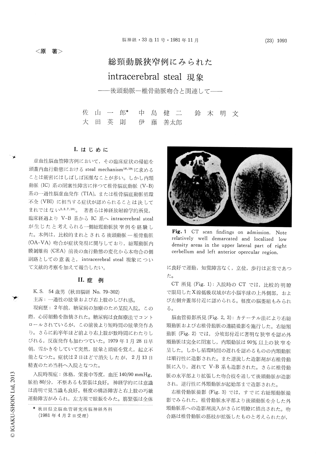
On admission slight dysarthria, skilled work dis-turbance in the right hand and nystagmus were the symptoms and signs which could be detected at that time. CT scan showed mild brain atrophy with well-demarcated and localized low density areas in the left anterior operculum and the right upper lateral part of the cerebellum. With Seldg-inger's method, a right common carotid angiography was performed, and it disclosed marked stenosis of the right common carotid artery, with an external carotid occlusion and over 90% stenosis of the in-ternal carotid artery. Furthermore, after perform-ing right vertebral angiography, a sufficient retro-grade filling of the external carotid territory was observed, via the well-developed occipital-vertebral anastomosis. As the diameter of the collateral channel was uniform, this might be termed a direct anastomosis.
Concerning the vertebro-basilar system, no occlu-sion or stenosis was detected. However, it was worthy to note that through the enlarged right posterior communicating artery, a blood stream was found running from the vertebro-basilar to the internal carotid system.
41 days after the last attack, a right carotid endarterectomy was performed and the postoperative course was uneventful.
On the postoperative angiography, the collateral channel via the occipital-vertebral anastomosis had almost disappeared and flow direction was reversed. The blood flow through the right posterior com-municating artery was also reversed, and by in-ternal carotid injection, opaque media flowed into the vertebro-basilar system. This patient was discharged without neurological deficits, and he has continued to be free from complaints.
In this case, ischemic events in the vertebro-basilar system might have been due to right com-mon carotid stenosis. There was a well-developed occipital-vertebral anastomosis as a collateral path-way to the occluded external carotid territory on the right side, and through the enlarged right posterior communicating artery, the flow was shifted from the vertebro-basilar to the right in-ternal carotid system. As a result of this re-distri-bution of blood flow, the secondary ischemic events might have appeared in the vertebro-basilar ter-ritory. And with a relative increase of vascular resistance, microemboli might lodge in the right superior cerebellar artery, a conclusion supported by CT scan. The pre- and postoperative changes of hemodynamics, as well as the postoperative course, suggest strongly that the preoperative is-chemic events in this patient might have been due to intracerebral steal phenomenon between the vertebro-basilar and the carotid system.

Copyright © 1981, Igaku-Shoin Ltd. All rights reserved.


