Japanese
English
- 有料閲覧
- Abstract 文献概要
- 1ページ目 Look Inside
I.はじめに
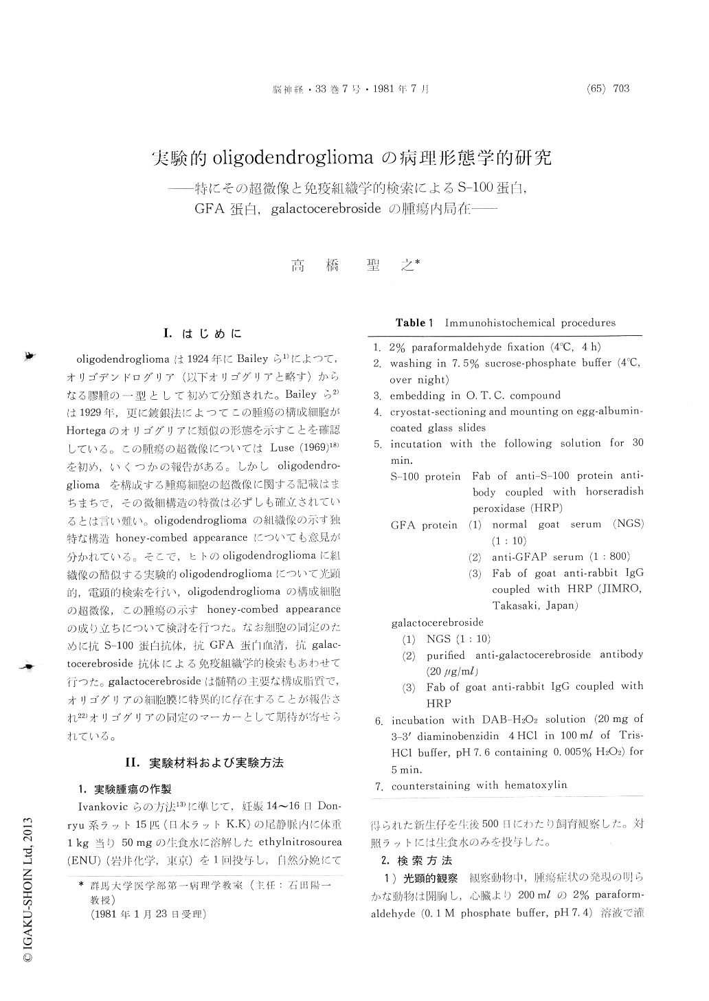
oligodendrogliomaは1924年にBaileyら1)によつて,オリゴデンドログリア(以下オリゴグリアと略す)からなる膠腫の一型として初めて分類された。Baileyら2)は1929年,更に鍍銀法によつてこの腫瘍の構成細胞がHortegaのオリゴグリアに類似の形態を示すことを確認している。この腫瘍の超微像についてはLuse (1969)18)を初め,いくつかの報告がある。しかしoligodendro—gliomaを構成する腫瘍細胞の超微像に関する記載はまちまちで,その微組構造の特徴は必ずしも確立されているとは言い難い。oligodendrogliomaの組織像の示す独特な構造honey-combed appearanceについても意見が分かれている。そこで,ヒトのoligodendrogliomaに組織像の酷似する実験的oligodendrogliomaについて光顕的,電顕的検索を行い,oligodendrogliomaの構成細胞の超微像,この腫瘍の示すhoney-combed appearanceの成り立ちについて検討を行つた。なお細胞の同定のために抗S−100蛋白抗体,抗GFA蛋白血清,抗galac—tocerebroside抗体による免疫組織学的検索もあわせて行つた。galactocerebrosideは髄鞘の主要な構成脂質で,オリゴグリアの細胞膜に特異的に存在することが報告され22)オリゴグリアの同定のマーカーとして期待が寄せられている。
Etylnitrosourea-induced experimental oligoden-drogliomas in rats were investigated with light and electron microscopy and by use of immuno-histochemical technique with antisera to S-100 and GFA proteins and anti-galactocerebroside antibody.
A total of 66 neuronal and nonneuronal tumors were produced in 44 litters treated with ethylnitro-sourea through the mothers. Of 66 tumors pro-duced 43 were found in the brain or spinal cord.
Histological classification of the glial tumors was made according to the cytology and pattern of predominant tumor cells. Tumors classified as oligodendroglioma amounted 26, of which 7 showed the histology typical of oligodendroglioma and 19 that of oligodendroglioma mixed with astrocytoma or with anaplastic glioma.
Localization of S-100 and GFA proteins in ex-perimental oligodendroglioma was studied immuno-histochemically. Astrocytic elements variously included in oligodendroglioma and mixed glioma showed positive brown staining for S-100 and GFA proteins. Anaplastic glia were also found weakly positive. The GFA protein was not deteced in oligdendroglioma tumor cells. Some of the oligo-dendroglioma cell nuclei were found to contain S-100 protein together with their narrow cytoplasm.
It was recently described that normal oligoden-droglia contained exclusively galactocerebroside. The cytological distribution of galactocerebroside was examined by immunoperoxidase method. In-terfascicular cells in the white matter showed positive staining. Galactocerebroside was also detected in normal ependymal cells and choroid plexus epithelium. Section of experimental oli-godendroglioma stained with antigalactocerebroside antibody disclosed the component cells to be negative.
Electron microscopic studies were made on a total 8 examples with oligodendroglial tumors. The neoplasms were mainly composed of loosely ar-ranged small dark cells that possessed very scanty cytoplasm and a few, delicate, short cytoplasmic processes. The cytoplasm of neoplastic cells appeared undifferenciated, but occasional cells con-tained microtubular structure about 25nm in width which was dispersed in the cytoplasm. The ex-tracellular space was abundant. Cells identifiedas astrocytic elements were found mixed in the tumor. The cells had longer and more prominient cell processes which contained bundles of glial filaments. Of 8 oligodendroglial tumors, 7 showed this type of the ultrastructure. In one case the tumor tissue which resembled a honeycomb by light microscopy was comprized of pale cells with an abundant cytoplasm. In a case examined astrocytic component cells showed a complex sheet-like processes were found around cell bodies myelinated fibers, and some unidentified cell processes.
The present investigation has established that the prevalent oligodendroglial cells in experimental oligodendroglioma are characterized in electron microscopy by their scanty cytoplasm with a few, short cytoplasmic processes. Microtubules were obserbed in some of the cells. The cells in oli-godendroglioma appear to preserve the ultrastruc-tural features described by Mori and Leblond for normal oligodendrocytes. It is also suggested in this study that the presence of an abundant extra-cellullar space probably acounts for the honey-combed structure in the light microscopy of ex-perimental oligodendroglioma.

Copyright © 1981, Igaku-Shoin Ltd. All rights reserved.


