Japanese
English
- 有料閲覧
- Abstract 文献概要
- 1ページ目 Look Inside
I.はじめに
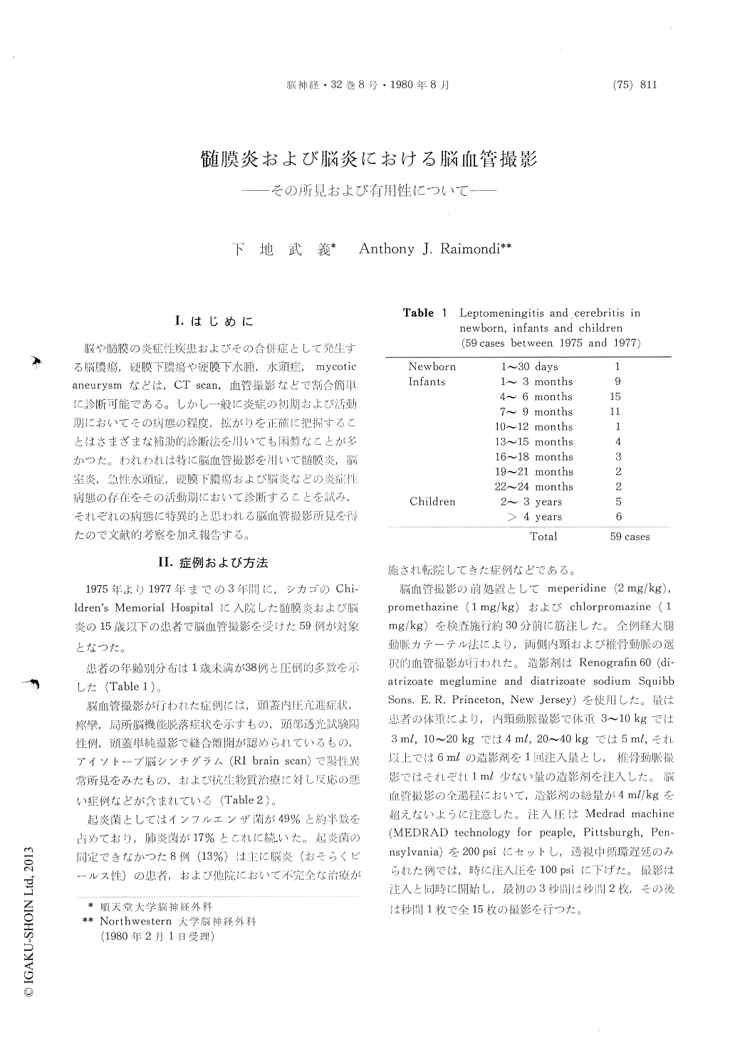
脳や髄膜の炎症性疾患およびその合併症として発生する脳膿瘍,硬膜下膿瘍や硬膜下水腫,水頭症,mycoticaneurysmなどは,CT scan,血管撮影などで割合簡単に診断可能である。しかし一般に炎症の初期および活動期においてその病態の程度,拡がりを正確に把握することはさまざまな補助的診断法を用いても困難なことが多かつた。われわれは特に脳血管撮影を用いて髄膜炎,脳室炎,急性水頭症,硬膜下膿瘍および脳炎などの炎症性病態の存在をその活動期において診断することを試み,それぞれの病態に特異的と思われる脳血管撮影所見を得たので文献的考察を加え報告する。
This is a report of the cerebral angiographic findings in cases of meningitis and cerebritis.
Fifty-nine patients, 38 of whom were under 1 year of age, underwent cerebral angiography by means of femoral catheterization. All the patients had signs of increased intracranial pressure, sei-zures, focal cerebral signs, positive transillumination of the head, and or abnormal brain scan findings. A few patients who did not respond to systemic antibiotics as was expected were also evaluated by means of cerebral angiography.
The causative organisms were Hemophilus in-fluenzae, pneumococcus, streptococcus, tuberculosis, E. coli, Pasteurella, and mycoplasma pneumoniae.
The following characteristic angiographic find-ings were observed in 18 cases of active meningitis :
(1) A hasy appearance around the arteries (halo formation) between the late arterial and capillary phases.
(2) Narrowing of the arteries in the basal cis-tern. This sometimes extended to the peripheral arteries.
(3) Irregular caliber following the narrowing of arteries (in few cases).
(4) Circulation time so slow that veins could beseen in the late arterial phase.
(5) Halo formation around the anterior chroidal artery and the clear appearance of the choroid plexus in the venous phase (when the infectious process reached the choroid plexus).
Six patients had acute hydrocephalus in addition to active meningitis.
Two patients were diagnosed as having subdural empyema with avascular space over the brain, an irregular appearance of the brain contour, and the above angiographic findings.
In the patients with subdural effusion and hy-drocephalus, which developed as complications of meningitis, no abnormal findings in the vessels were observed.
Cerebritis could be identified on the angiograms by two signs:
(1) local swelling of the brain (mainly the tem-poral lobe) and
(2) staining around the veins without any ab-normal signs in the arterial phase (laminar stain-ing).
In our opinion, halo formation in meningitis and laminar staining in cerebritis may result from leakage of the contrast medium through the broken blood brain barrier, as well as hyperemia due to the inflammatory process.
In conclusion, angiography is a meaningful test by which to determine the phase of meningitis and cerebritis. These two conditions should be treated based on valid information obtained by means of CSF examinations and neuroradiological tests, es-pecially CT scan and cerebral angiography.

Copyright © 1980, Igaku-Shoin Ltd. All rights reserved.


