Japanese
English
- 有料閲覧
- Abstract 文献概要
- 1ページ目 Look Inside
I.はじめに
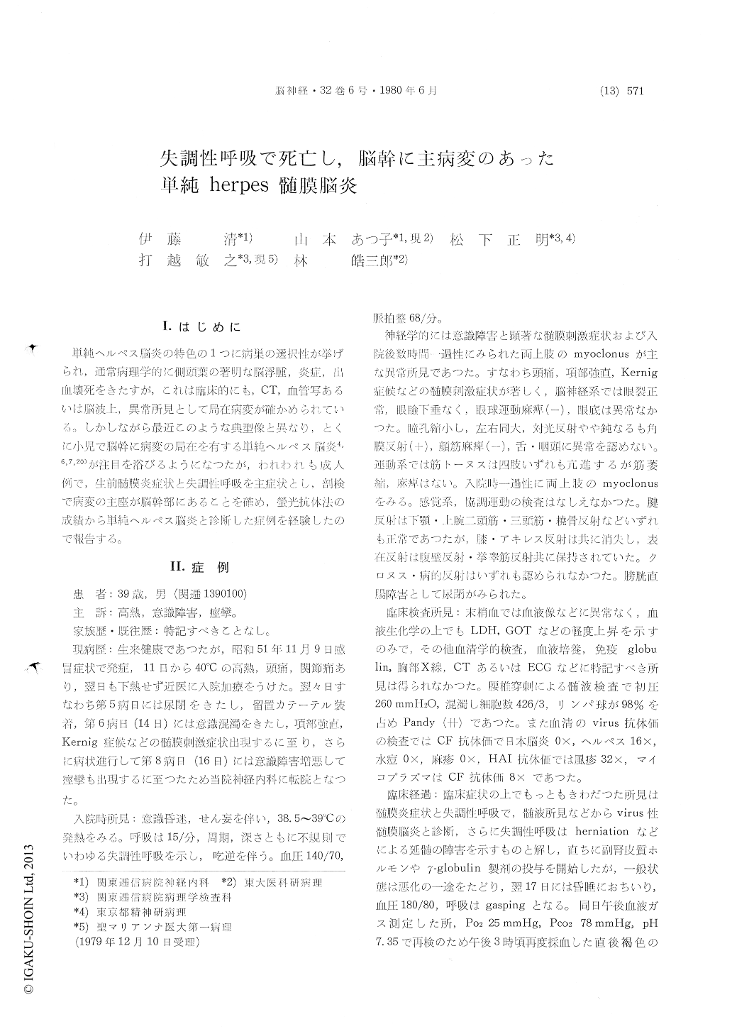
単純ヘルペス脳炎の特色の1つに病巣の選択性が挙げられ,通常病理学的に側頭葉の著明な脳浮腫,炎症,出血壊死をきたすが,これは臨床的にも,CT,面管写あるいは脳波上,異常所見として局在病変が確かめられている。しかしながら最近このような典型像と異なり,とくに小児で脳幹に病変の局在を有する単純ヘルペス脳炎4,6,7,20)が注目を浴びるようになつたが,われわれも成人例で,生前髄膜炎症状と失調性呼吸を主症状とし,剖検で病変の主座が脳幹部にあることを確め,螢光抗体法の成績から単純ヘルペス脳炎と診断した症例を経験したので報告する。
It has been commonly accepted that herpes sim-plex encephalitis does not present as brainstem disorder but temporal lobe necrosis. However, re-cently a few case reports of unusual brainstem encephalitis caused by herpesvirus hominis, especi-ally in the childhood, called attention to the pos-sibility of this disease.
In this paper we represent atypical case at ne-cropsy findings died of acute respiratory failure in the adult.
A 39-year-old man, who had been well before 7 days prior to admission, was acutely ill with fever like common cold and the othe symptoms such as urinary incontinence, stiffness of the neck and con-vulsion were manifested. On admission, he was stuporous and had high fever and irregular non-periodical breathing of various amplitude (ataxic breathing). Neurological examination revealed the following abnormal findings: remarkable meningeal signs, miotic pupil, myoclonic movement of both hands, areflexia of knee jerk and urinary inconti-nence.
Laboratory studies showed as follows: serum complement-fixing antibody titre to herpes virus rose 1: 16 on the day of admission. Cerebrospinal fluid revealed moderate pleocytosis and slight ele-vation of protein content. Neither of viruses had been recovered from the cerebrospinal fluid.
He became rappidly deteriorated and died of respiratory failure on the next day of admission.
Autopsy revealed grossly hyperemic meninges, severe cerebral edema and marked uncal and tonsillar herniations. Microscopical examination showed diffuse lymphocytic meningoencephalitis. Pronounced infiltrative changes mainly consisting of lymphocytes, plasma cells and histiocytes were found extensively in the meninges and the peri-vascular spaces. However the encephalitic lesions were not observed in the both cerebral hemispheres, those were localized mainly in the brainstem. The perivascular cuffing, microglial proliferation, glial nodule, neuronophagia, nerve cell necrosis were scattered throughout the brainstem, involving sub-stantia nigra, pontine tegmentum, pontine base, cerebellar cortex, mostly severe in the medulla oblongata. A few inclusion body like structure were seen. Cryostat sections from front-temporal cortex were examined by the direct immunofluo-rescent technique, and the herpes virus antigens were observed in the glial cells and arachnoidal membranes.
These clinicopathological studies lead to the fol-lowing conclusions : this case is diagnosed herpes simplex miningo-encephalitis being dead of acuterespiratory failure, that is ataxic breathing, and the main lesion is distributed in the brainstem, particularly in the medulla oblongata. The ataxic breathing, that is frequently observed on the ex-perimental study of the brain section at the level of the medulla,seems to be ascribed to the medul-lary lesion of this case.
Brainstem encephalitis is an ill-defined entityand can not usually attributed to herpes simplex infection, but there are 6 case reports of such un-usual cases. Most reported cases are infant or child and those cases have mostly gravy symptomes with benign prognosis, but the respiratory dis-turbances were frequently noted as well as in this case during acute and recovery stages.

Copyright © 1980, Igaku-Shoin Ltd. All rights reserved.


