Japanese
English
- 有料閲覧
- Abstract 文献概要
- 1ページ目 Look Inside
I.はじめに
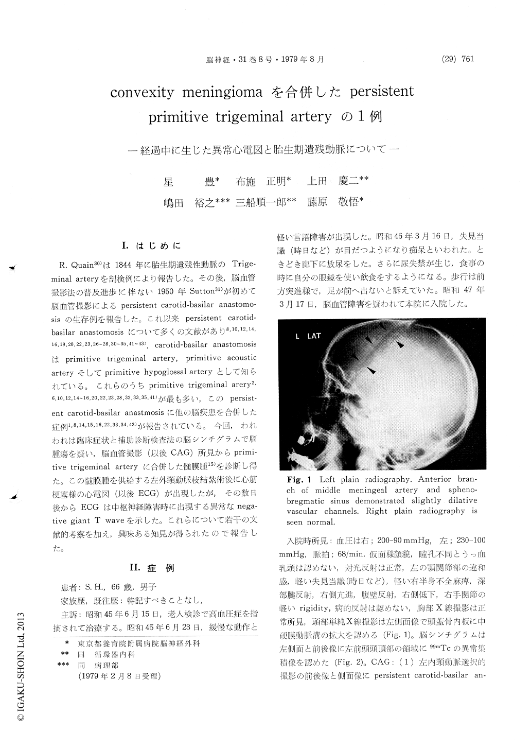
R. Quain30)は1844年に胎生期遺残性動脈のTrige—minal arteryを剖検例により報告した。その後,脳血管撮影法の普及進歩に伴ない1950年Sutton31)が初めて脳血管撮影によるpersistent carotid-basilar anastomo—sisの生存例を報告した。これ以来persistent carotid—basilar anastomosisについて多くの文献があり8,10,12,14,16,18,20,22,23,26〜28,30〜35,41〜43),carotid-basilar anastomosisはprimitive trigeminal artery,primitive acousticarteryそしてprimitive hypoglossal arteryとして知られている。これらのうちprirnitive trigeminal arery2,6,10,12,14〜16,20,22,23,28,32,33,35,41)が最も多い,このpersist—ent carotid-basilar anastmosisに他の脳疾患を合併した症例1,8,14,15,16,22,33,34,43)が報告されている。今回,われわれは臨床症状と補助診断検査法の脳シンチグラムで脳腫瘍を疑い,脳血管撮影(以後CAG)所見からprimi—tive trigeminal arteryに合併した髄膜腫15)を診断し得た。この髄膜腫を供給する左外頸動脈枝結紮術後に心筋梗塞様の心電図(以後ECG)が出現したが,その数日後からECGは中枢神経障害時に出現する異常なnega—tive giant T waveを示した。これらについて若干の文献的考察を加え,興味ある知見が得られたので報告した。
A case of 66 years old man of persistent primitive trigeminal artery with convexity meningioma is reported. This patient was admitted to the Yoikuen Hospital on March 17, 1972, because of a complaint of progressive dysarthria, incontinentia urinaly, disorientation, and slight right hemiparesis with propulsive gait. Two years earlier, this patient had arised progressive disturbance of speech with hypertension, but there was not post history of neurological, cardiac disease and abnormal record of ECG. On admission, he was dis-oriented, and had disturbance of sensory at left maxial joint and slight right hemiparesis. Blood pressure was 200/ 90 mmHg at right, 230/100 mmHg at left side. Pulse rate was 68 per minute. He had not papil-ledema and anisocoria. A lumber puncture revealed clear spinal fluid at an openning of 160 mmH2O. Hematological findings and blood chemistry were showen in Table 1, 2.
Brain scans obtained increased radioactivity in peripheral of left cerebral hemisphere on anterior and posterior views. A mass lesion in convexity region on the left lateral view was suggested by brain scan, and selective external carotid angiogram. Selective internal carotid angiogram faild to show a posterior communicating, but this angiogram demonstrated an anomalous anastomosis betweenthe internal carotid artery and the basilar artery. Superior cerebeller and posterior cerebral arteries were visible. This arterioangiogram is the carotid-basilar anastomosis which is called persistent primitive trigeminal artery. Contrary to arterio-venous fistula which should be increased, it is said that carotid-basilar anastomosis of cerebral blood flow (CBF) is in the range of normal values. Ligation of a branch of left external carotid artery may produce alterations in electrocardiogram (ECG), deep inverted T wave, elevated ST in V2, V3, appeared Q wave in II, III, aVf, V3-6, negative T wave in I, II, III, aVf, V3-6. And QT time was O.56 sec. This record of ECG is probable acute myocard infarction. Since then this ECG record had demonstrated the decraesed bight of R and deep negative T wave. This deep negative gaint T wave is ECG record of disturbance of central nervous system.
This patient demonstrated all of those ECG changes, and ECGs, recorded after external carotid artery ligation resembled ECG of acute subendo-cardial infarction and disturbance of NCS. Ab-normal ECG record in post operation of external carotid artery demonstrated ischemic-like electro-cardiographic disturbance. The reason for this may have been related to the long term of com-pression at surface of brain cortex by meningioma. It seems likely that the abnormal ECGs, to be seen particulary in this case with primitive trigeminal artery and meningioma, resulted from a combination of 2 factors. Stimulation of autonomic nerves controlling the circle Willis, transient brain damage owing to blood, and ischemia from hypo-circle of deep perforating vessels may have contributed to abnormal ECG in this patient. Stimulation of autonomic nerves by operation arised from auto-nomic stimulation of hals to central nerves, and it seemed that vasocardiac reflex arising in circle Willis was possible factor which caused ECG changes.
Copyright © 1979, Igaku-Shoin Ltd. All rights reserved.