Japanese
English
- 有料閲覧
- Abstract 文献概要
- 1ページ目 Look Inside
I.はじめに
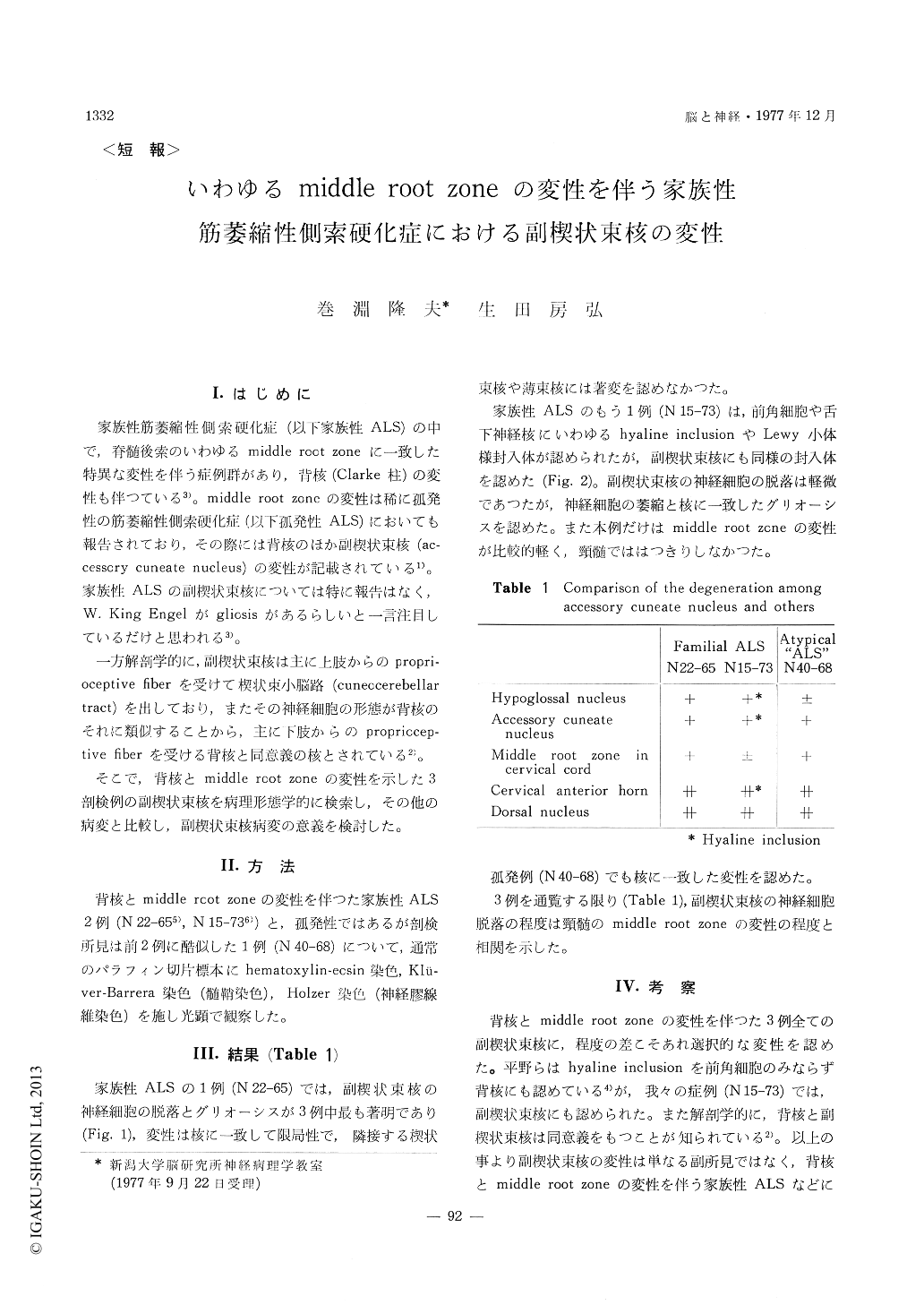
家族性筋萎縮性側索硬化症(以下家族性ALS)の中で,脊髄後索のいわゆるmiddle root zoneに一致した特異な変性を伴う症例群があり,背核(Clarke柱)の変性も伴つている3)。middle root zoneの変性は稀に孤発性の筋萎縮性側索硬化症(以下孤発性ALS)においても報告されており,その際には背核のほか副襖状束核(ac—cessory cuneate nucleus)の変性が記載されている1)。家族性ALSの副楔状東核については特に報告はなく,W.KingEngelがgliosisがあるらしいと一言注目しているだけと思われる3)。
一方解剖学的に,副楔状束核は主に上肢からのpropri—oceptive flberを受けて楔状束小脳路(cuneocerebellartract)を出しており,またその神経細胞の形態が背核のそれに類似することから,主に下肢からのpropriccep—tive flberを受ける背核と同意義の核とされている2)。
It was reported that some cases of familial ALS showed the unique degeneration corresponding to the so-called "middle root zone" of the spinal posterior funiculus and degeneration of the dorsal nucleus. In a few cases of atypical sporadic ALS, the degen-eration of middle root zone was also reported, and besides degeneration was described in the accessory cuneate nucleus as well as in the dorsal nucleus.
Anatomically, accessory cuneate nucleus is consid-ered to be the medullary equivalent of dorsal nucleus due to the facts that the accessory cuneate nucleus receives proprioceptive fibers mostly from upper limb and gives rise to cuneocerebellar tract and that neurons of the accessory cuneate nucleus and the dorsal nucleus are morphologically similar.
We examined the accessory cuneate nucleus light-microscopically in two cases with familial ALS and one with sporadic ALS all showing degeneration of dorsal nucleus and middle root zone.
All the three cases showed certain degeneration of accessory cuneate nucleus such as atrophy and depopulation of neurons and gliosis. In one case with familial ALS, the anterior horn cells contained intracytoplasmic hyaline inclusions, and the similar inclusions were also observed in the accessory cuneate nucleus.
No significant change was found in the cuneate and gracil nuclei in three cases.
Those suggest that the degeneration of the ac-cessory cuneate nucleus is not an incidental but a fundamentally analogous finding to the degeneration of the dorsal nucleus in familial ALS and possibly a primary alteration as a part of motor neuron disease.
In the three cases examined, there is a correlation between the neuronal depopulation of accessory cuneate nucleus and the degeneration of middle root zone in the cervical cord. To understand the degeneration of the middle root zone of the cervical cord, which does not have dorsal nucleus, it is proper to take into consideration the degeneration of the accessory cuneate nucleus in place of the dorsal nucleus.
The significance of the degeneration of accessory cuneate nucleus will be its anatomical location ; since the dorsal nucleus is located near the anterior horn, while the accessory cuneate nucleus is far from it and even from the hypoglossal nucleus. The nucleus may make an important key to the pathogenesis of motor neuron disease.

Copyright © 1977, Igaku-Shoin Ltd. All rights reserved.


