Japanese
English
- 有料閲覧
- Abstract 文献概要
- 1ページ目 Look Inside
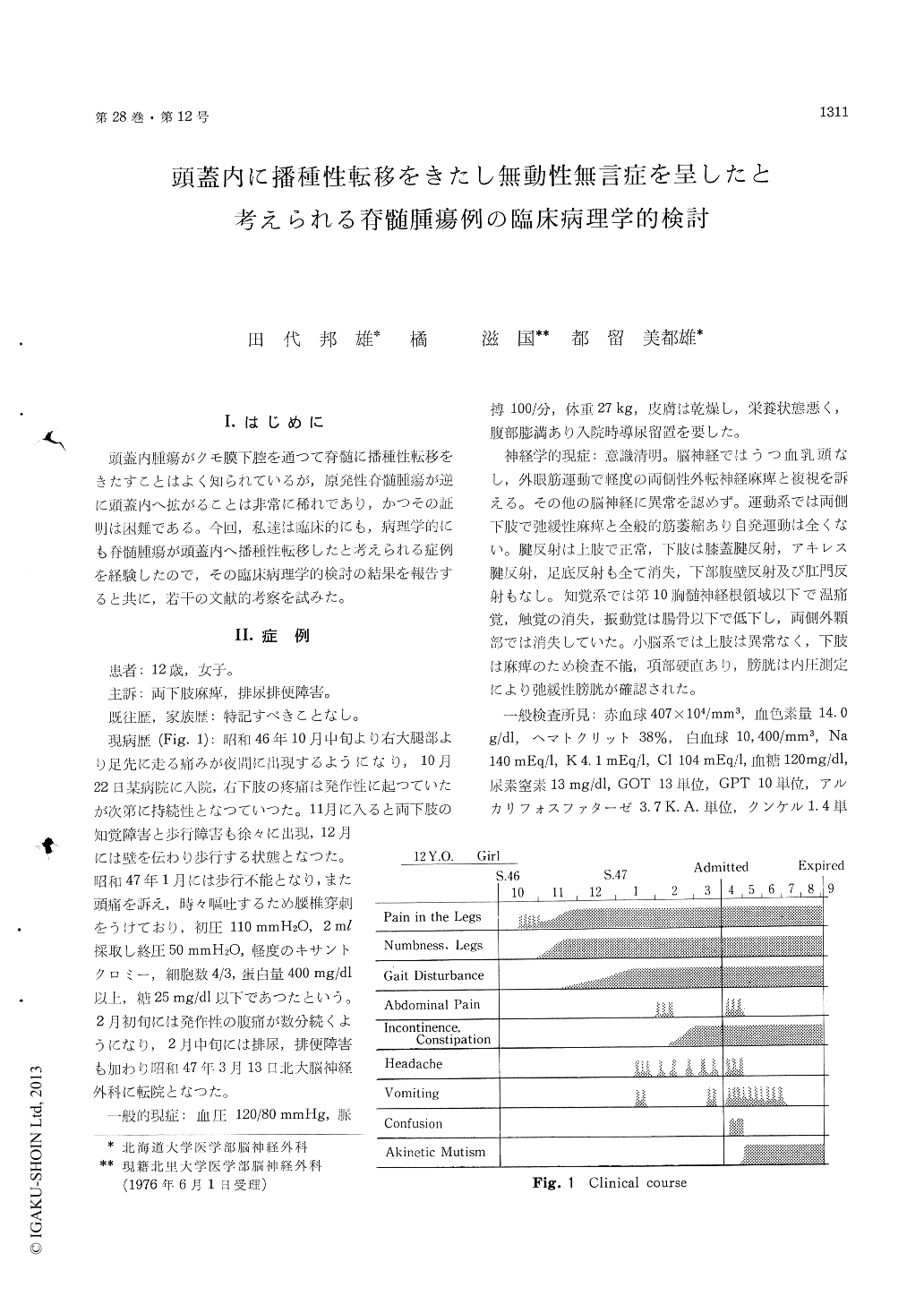
I.はじめに
頭蓋内腫瘍がクモ膜下腔を通って脊髄に播種性転移をきたすことはよく知られているが,原発性脊髄腫瘍が逆に頭蓋内へ拡がることは非常に稀れであり,かつその証明は困難である。今回,私達は臨床的にも,病理学的にも脊髄腫瘍が頭蓋内へ播種性転移したと考えられる症例を経験したので,その臨床病理学的検討の結果を報告すると共に,若干の文献的考察を試みた。
Intracranial dissemination of primary spinal cord tumors is an extremely rare occurrence.
A 12-year-old girl first developed nocturnal pain in the right leg 5 months prior to admission, follow-ed fairly rapidly by constant pain, weakness and numbness of both legs, and difficulty in walking. She was totally bed-ridden by 2 months prior to admission, and also complained of occasional ab-dominal pain, headache and vomiting in addition to urinary incontinence. She was admitted to Hokkaido University Hospital on March 13, 1972.
Neurological examination revealed minimal bi-lateral abducens palsy, flaccid paraplegia, absent DTRs and plantar responses in both legs, and sensory loss below D10. The lower abdominal reflexes were also absent. Cisternal myelogram disclosed total subarachnoid block at the level of Th10-11 (Fig. 2), and intramedullary tumor involving from conus medullaris to dorsal cord was partially re-moved by laminectomy. Approximately 3 weeks following operation, she started to have signs of increased intracranial pressure, for which ventricular drainage was performed. She became gradually in the state of akinetic mutism, and expired on September 2, 1972 in extremely poor general con-dition.
Autopsy disclosed a huge spinal cord tumor in-volving from conus medullaris to dorsal cord (Fig. 4a), which was glioblastoma multiforme histologi-cally (Fig, 7). The subarachnoid space at the base of the brain was filled with thick tumor infiltration (Fig. 4b).
Coronal sections of cerebral hemispheres and transverse sections of brain stem and cerebellum clealy showed extensive tumor growth along the ventricle system and subarachnoid space with defi-nite intramedullary infiltration (Fig. 5 & 6). The intracranial tumors were identical, being astro-cytoma.
The clinical course and neuropathological findings in this case made us believe that primary neoplastic changes occurred in the spinal cord with subsequent intracranial dissemination.
Review of the literatures revealed only 9 autopsy proved cases of intracranial dissemination of spinal tumors, to which our case was added (Table 1).
Copyright © 1976, Igaku-Shoin Ltd. All rights reserved.