Japanese
English
- 有料閲覧
- Abstract 文献概要
- 1ページ目 Look Inside
I.はじめに
高血圧性脳内出血その他,各種の脳内血腫において,その血腫の拡がりと個々の神経核,神経索,脳室との関係等を把握することは,その機能予後を推定する上に重要なことであるが,これまでの脳血管撮影,脳室造影等のみでは,極めて困難であつた。この問題が近い将来,C.A.T.の一般的普及と共に,大幅に解決される見通しが立つたこと2,11)は喜ばしいことであるが,我々はこれまで約三年間,開頭血腫別除術に際し,ごく少量のPantopaqueを使用することにより,上記目的をほぼ達することが出来たので,術後のStereotaxic evaiuationの方法を含めて,ここに報告する。
これまでにこの造影法を行った症例は外側型脳内血種55例,特発性脳内血腫9例,脳橋出血3例,外傷性脳内出血4例,脳動脈瘤及び脳動静脈奇型に伴う脳内血腫5例の計76例である。
In order to confirm the depth and extent of thehematoma cavity in various types of intracerebralhematoma, small amount of Pantopaque was left inthe cavity at the time of craniotomy as the follow-ing method. Small pieces of cottonoid oxycell weresoaked in Pantopaque and were fixed on the wallof the hematoma cavity on such three spots as inthe most anterior portion, in the most posteriorportion and in the deepest portion after evacuationof hematoma.
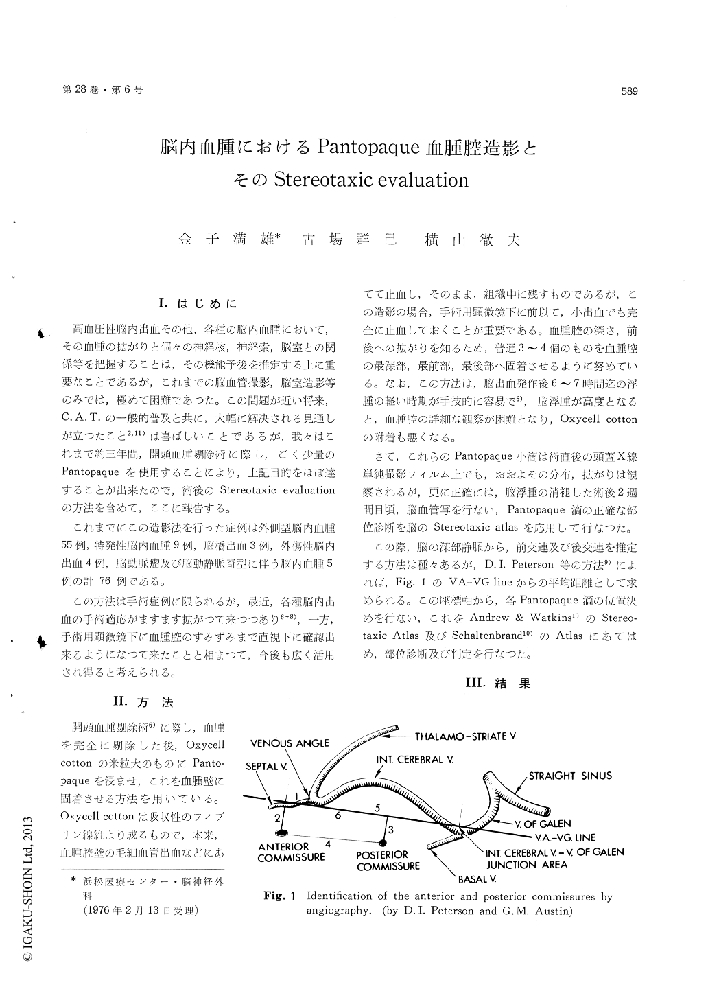
Those few drops of Pantopaque were read onpastoperative carotid angiogram and spotted on thestereotaxic atlas of Schaltenbrand or of Andrewsand Watkins. On this measurement, anterior andposterior commissures were identified on venousphase of carotid angiography by the method of D. I.Peterson and G. M. Austin.
This Pantopaque radiography was applied on 76cases of several types of intracerebral hematomaand it was much valuable to estimate the correlationbetween the hematoma and the surrounding cereb-ral tissue such as basal ganglia, internal capsule,lateral ventricle and so on and thus to assess theprognosis of individual case.

Copyright © 1976, Igaku-Shoin Ltd. All rights reserved.


