Japanese
English
- 有料閲覧
- Abstract 文献概要
- 1ページ目 Look Inside
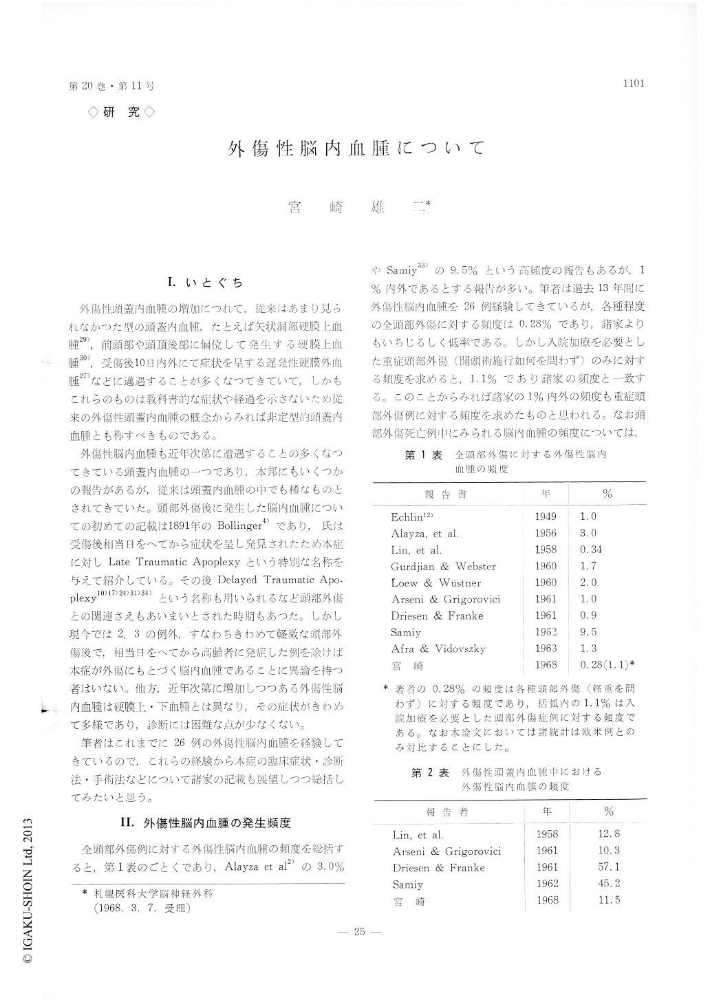
I.いとぐち
外傷性頭蓋内血腫の増加につれて,従来はあまり見られなかつた型の頭蓋内血腫,たとえば矢状洞部硬膜上血腫29),前頭部や頭頂後部に偏位して発生する硬膜上血腫30),受傷後10日内外にて症状を呈する遅発性硬膜外血腫27)などに邁遇することが多くなつてきていて,しかもこれらのものは教科書的な症状や経過を示さないため従来の外傷性頭蓋内血腫の概念からみれば非定型的頭蓋内血腫とも称すべきものである。
外傷性脳内血腫も近年次第に遭遇することの多くなつてきている頭蓋内血腫の一つであり,本邦にもいくつかの報告があるが,従来は頭蓋内血肺の中でも稀なものとされてきていた。頭部外傷後に発生した脳内血腫についての初めての記載は1891年のBollinger4)であり,氏は受傷後相当日をへてから症状を呈し発見されたため本症に対しLate Traumatic Apoplexyという特別な名称を与えて紹介している。その後Delayed Traumatic Apo—plexy10)17)24)31)34)という名称も用いられるなど頭部外傷との関連さえもあいまいとされた時期もあつた。しかし現今では2,3の例外,すなわちきわめて軽微な頭部外傷後で,相当日をへてから高齢者に発症した例を除けば本症が外傷にもとづく脳内血種であることに異論を持つ者はいない。他方,近年次第に増加しつつある外傷性脳内血腫は硬膜上・下血腫とは異なり,その症状がきわめて多様であり,診断には困難な点が少なくない。
Although the epidural and subdural hematomas caused by craniocerebral injury have been investi-gated at considerable length, relatively few papers have been devoted to traumatic intracerebral hema-toma.
The traumatic intracerebral hematoma have been described as uncommon lesion caused by head injury. In recently, due to increased number of traffic and industrial accident, the traumatic intracerebral hema-toma is probably more common than is indicated by literature.
The purpose of the present report are : (1) to em-phasize the importance of recognizing traumatic intracerebral hematoma, (2) to review the clinical course and symptom, (3) to present important points to make a diagnosis, particularly important evidence of cerebral angiogram, (4) to review the surgical operative procedure- and, (5) to evaluate the prog-nosis of surgical evacuation, by authors own experi-ences of 26 cases and literatures.
The traumatic intracerebral hematoma divided into three types by the time appeared symptom after head injury, type I is that symptom appeared within two days after head injury, type II is that five days and type III is that ten days. The frequency is 46.1%, 30.8% and 23.1%. The symptoms are differ-ent in these three types. The major cases of type I are lowering of conciousness level after interval and other cases are continuous state of somnolence and delirium or focal cerebral symptom. The type II are somnolence or delirium after interval, or focal cerebral symptom meantime the consciousness level getting up. The type III are change of mental state or focal cerebral symptom under clear con-sciousness.
The angiographic evidence of traumatic intra-cerebral hematoma in frotal lobe is quite same with the tumor in this site. The intracerebral hematoma in temporal lobe shows upward dislocation of middle cerebral artery with or without shifting of anterior cerebral artery to opposite side. The angiographic diagnosis of intracerebral hematoma in temporal lobe have to done carefully, because that hematoma has few or no focal cerebral symptom and sign and has no shifting of anterior cerebral artery in three fifth.
Copyright © 1968, Igaku-Shoin Ltd. All rights reserved.