Japanese
English
- 有料閲覧
- Abstract 文献概要
- 1ページ目 Look Inside
I.はじめに
焦点性てんかんにおける脳循環の増加については,すでに諸家により報告されている6,2,7,11)。しかし,発作期と発作間激期の脳循環の変化については,不明な点が少なくない。
Ingvar3)は133Xe clearance法による局所脳循環(以下rCBFと略す)測定装置を使用し,視力障害を伴う後大脳動脈閉塞症例の後頭葉焦点部位にrCBFの大きな増加を認め報告した。しかし,これまで焦点性てんかんの発作期と発作間歓期のrCBFの比較,さらにrCBFよりみた焦点部位の病態生理に関する報告は稀である。
Regional cerebral blood flow (rCBF) was measured in 10 patients with focal cortical epilepsy using the the 254 Detector Dynamic Gamma Camera by the Xe-133 intracarotid method. Electroencephalo gram, mean arterial blood pressure and pCO2 were measured also during the rCBF study.
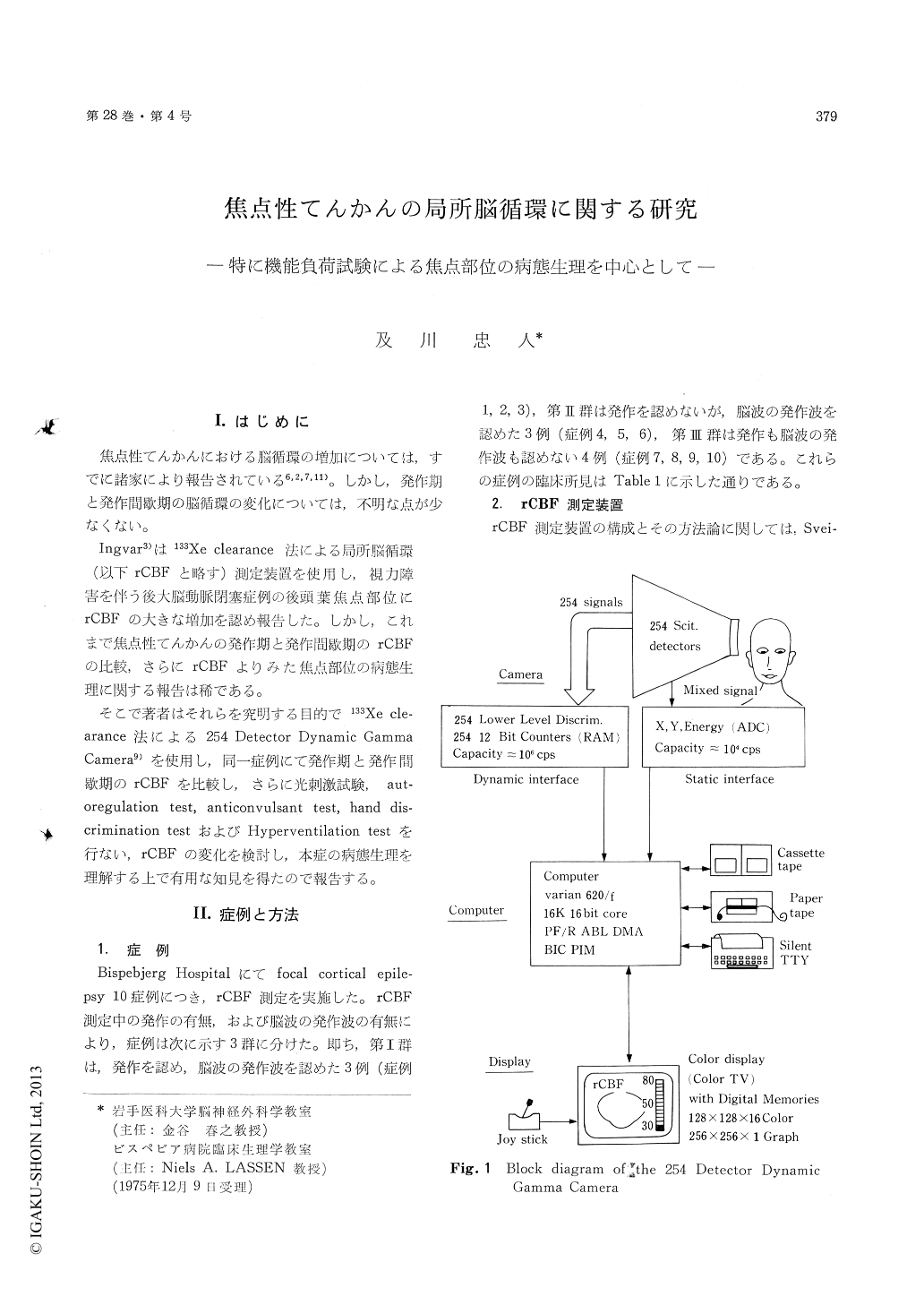
The 254-detector system is coupled on line to a small computer and results were obtained practically with no delay only 150 sec. after injection of Xe-133 on a color television. The system of display can show rCBF patterns and patient identification via a digital memory containing 128 × 128 16-level cells for color display and 256 × 256 cells in black and white.
Various functional tests e. g. photic stimulation test, anticonvulsant injection test, autoregulation test with angiotensin and hand discrimination test were performed for the purpose of observing changes of rCBF patterns during these tests.
The results obtained in the study were as follows :
1) The focus in the patient with hemi-epilepsy (case 1) showed from twice to ten times the rCBF of the non-focal areas. Two foci with increased rCBF were confirmed in two patients (case 2 and 3) during their respective epileptic seizures.
2) Even patients with no abnormalities of rCBF during their respective interictal phases demon-strated increased rCBF following photic stimulation. However, the patients who had already increased rCBF during their respective interictal phases did not reveal any signs of increased rCBF in all the areas including the focus.
3) Following anticonvalsant injection in six patients, five of the patients demonstrated markedly decreased rCBF in both their respective focal hy-peremic areas and the non-affected areas of the whole hemisphere, whereas the remaining one re-vealed no signs of changes in rCBF
4) Following angiotensin injection in six patients, while three of the patients demonstrated increased. CBF in their respective focal hyperemic areas, one of the remaining psatient who was a patient with postoperative megingioma revealed decreased rCBF and another two revealed no changes of rCBF.
5) All of the four patients given the hand dis-crimination test demonstrated increased rCBF in the sensorimotor area.
6) While the electroencephalogram of the patient with postoperative megingioma revealed no patho-logic signs in the mirror focus of the opposite side, focal hyperemic area with markedly increased rCBF was elicited by use of photic stimulation.

Copyright © 1976, Igaku-Shoin Ltd. All rights reserved.


