Japanese
English
- 有料閲覧
- Abstract 文献概要
- 1ページ目 Look Inside
I.はじめに
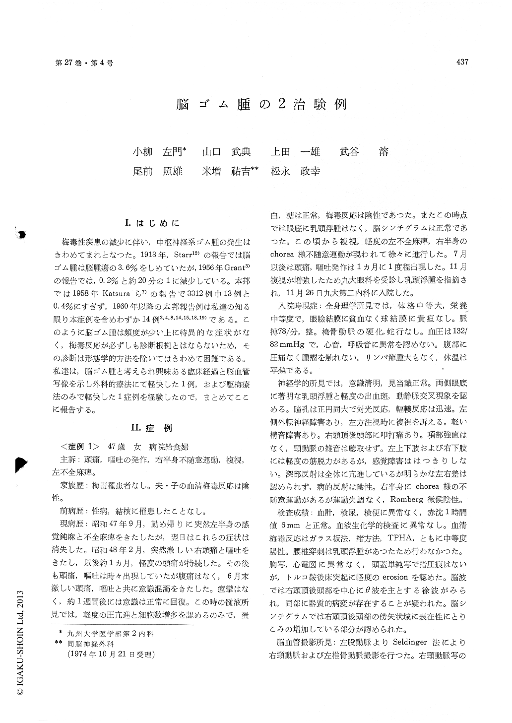
梅毒性疾患の減少に伴い,中枢神経系ゴム腫の発生はきわめてまれとなつた。1913年,Starr12)の報告では脳ゴム腫は脳腫瘍の3.6%をしめていたが,1956年Grant3)の報告では,0.2%と約20分の1に減少している。本邦では1958年Katsuraら7)の報告で3312例中13例と0.4%にすぎず,1960年以降の本邦報告例は私達の知る限り本症例を含めわずか14例2,4,8,14,15,18,19)である。このように脳ゴム腫は頻度が少い上に特異的な症状がなく,梅毒反応が必ずしも診断根拠とはならないため,その診断は形態学的方法を除いてはきわめて困難である。私達は,脳ゴム腫と考えられ興味ある臨床経過と脳血管写像を示し外科的療法にて軽快した1例,および駆梅療法のみで軽快した1症例を経験したので,まとめてここに報告する。
Two cases of intracranial gumma, the one surgi-cally and the other medically treated, are presentedin this report.
Case 1; A 47-year-old female was admitted be-cause of increasing visual disturbance and right-sided involuntary movement. Twelve months priorto admission she had experienced transient lefthemiparesis and numbness, which completely re-covered within 24 hours. Occasional headache andvomiting appeared 4 months after the initial attack,followed by an episode of disturbance of cons-ciousness lasting for about one week. Left hemi-paresis, double vision and involuntary movementon the right side of the body gradually developedduring the last several months. A fundoscopicexamination revealed papilledema, and serologicaltests for syphilis were positive. Spinal tap wasnot performed because of the presence of increasedintracranial pressure. Cerebral angiograms showedsigns of an intracranial space occupying lesionwith localized narrowing and irregularity of arteriallumen.
At surgery an intracranial granuloma firmly adh-eredto the thickned dura was found in the rightparietal parasagittal region. Histological exami-nation of the surgical specimens disclosed a syphiliticgranuloma.
She was vigorously treated with penicillin fortwo weeks after surgery and discharged with amoderate left hemiparesis as a neurological deficit.
Case 2; The patient was 65-year-old male, whohad been complaining of persistent headach for onemonth. He was admitted because of disturbanceof consciousness.
On admission he was stuporous and the neuro-logical examination disclosed left hemiparesis andequivocal early papilledema. Serological tests forsyphilis were positive in both the blood and thecerebrospinal fluid. Examination of the CSF re-vealed a pleocytosis and an increase of total protein.
An elevation of the right middle cerebral arterygroup with an equivocal shift of subcallosal portionof the right anterior cerebral artery was demon-strated by cerebral angiography, suggesting thepresence of a mass lesion in the right temporallobe. He was treated with penicillin (600,000unit, i. m.) and bithmuth bisalycilate (1.5g, i. v.)for eight weeks. The level of consciousness beganto improve within a couple of days after the treat-ment was started, and the neurological abnormalitieswere completely cleared at the end of two months,when the repeated angiography and serological testsfor syphilis of the CSF were also negative.
It was emphasized in this report that a possibilityof intracranial gumma should always be consideredin cases with intracranial mass lesion with positiveserological tests for syphilis in the blood, althoughthe incidence of neurosyphilis has recently beendecreased. A medical treatment should be triedbefore surgical approach when intracranial gummais strongly suggested, and the patient's condition isjustifiable for it.

Copyright © 1975, Igaku-Shoin Ltd. All rights reserved.


