Japanese
English
- 有料閲覧
- Abstract 文献概要
- 1ページ目 Look Inside
はじめに
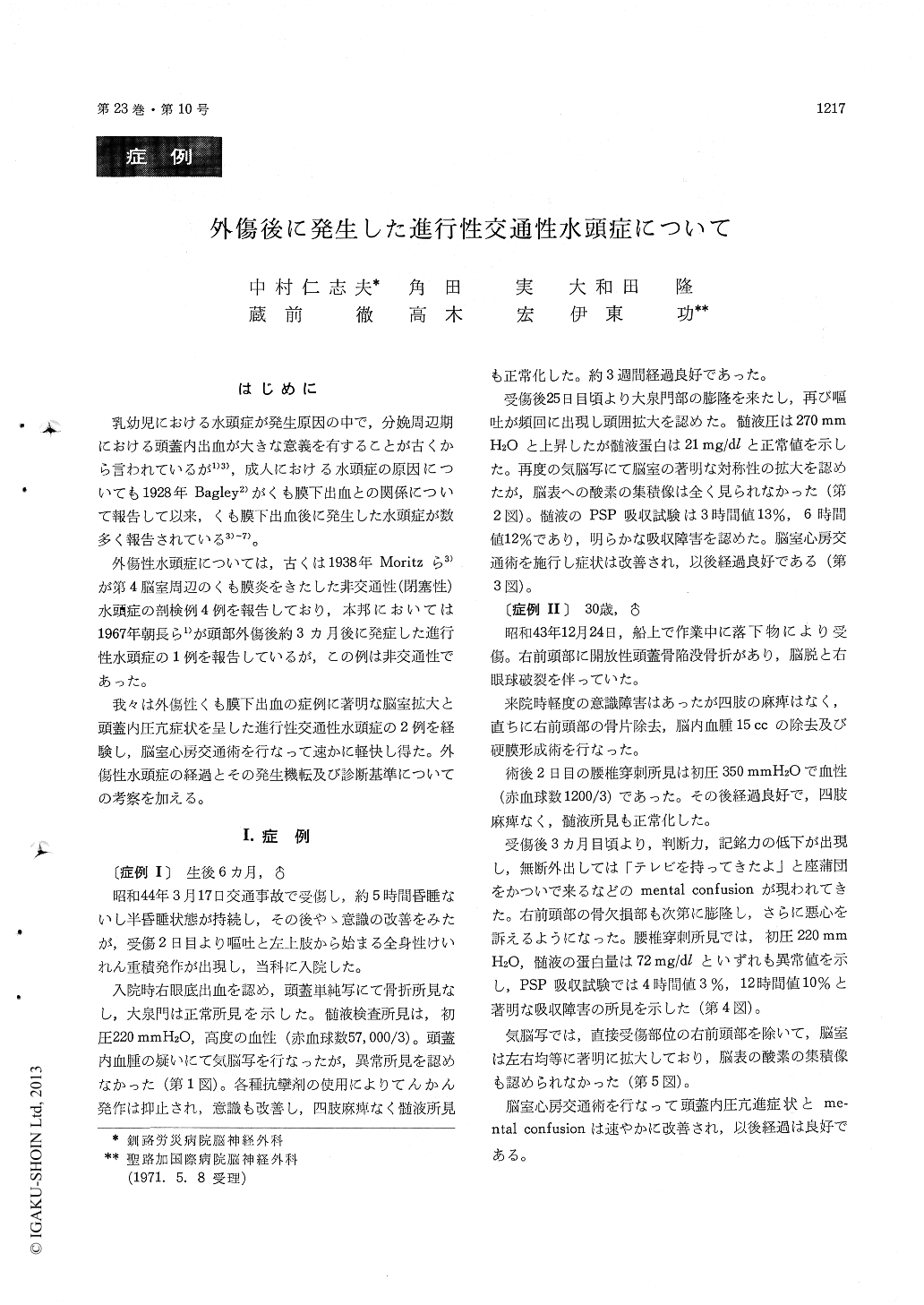
乳幼児における水頭症が発生原因の中で,分娩周辺期における頭蓋内出血が大きな意義を有することが古くから言われているが1)3),成人における水頭症の原因についても1928年Bagley2)がくも膜下出血との関係について報告して以来,くも膜下出血後に発生した水頭症が数多く報告されている3)〜7)。
外傷性水頭症については,古くは1938年Moritzら3)が第4脳室周辺のくも膜炎をきたした非交通性(閉塞性)水頭症の剖検例4例を報告しており,本邦においては1967年朝長ら1)が頭部外傷後約3カ月後に発症した進行性水頭症の1例を報告しているが,この例は非交通性であった。
Previous reports concerning the causes of hydro-cephalus have suggested that one of the prodispos-ing factors might be the impairment of the CSF absorption mechanism following subarachnoid he-morrage in the head injury or ruptured aneurysm. We present two cases of progressive non-obstrutive hydrocephalus supposed to be due to the head injuries.
Case I. A baby aged 6 months was injured by a traffic accident on 17th of March, 1969. The next day he was transferred to our hospital with vomit-ting and status epilepticus. Physical and neurologi-cal examinations revealed negative except for the hemorrhage in right optic fundus. Lumbar punc-ture revealed bloody liquor under normal pressure. PEG showed no abnormality at that time. The clinical course was uneventful until 3 weeks after the head injury, when the patient began to be suffered from recurrent vomitting accompanied with swelling on the great fontanelle and enlargement of the skull. The CSF pressure increased up to 270 mm H2O. Intrathecal PSP absorption test revealed 25% at 6 hours. PEG showed marked symmetrical dilatation of all ventriclar system and no subarac-hnoid air over the hemispheres. The patient reco-vered good health after the V-A shunt operation.
Case II. A workman aged 30 years with right frontal compound skull fracture was admitted to our hospital on 24th of December, 1968. His con-sciousness was a little confused but showed no other neurological manifestations when urgent operation was performed immediately. Lumbar puncture revealed bloody liquor under the pressure of 350 mm H2O two days after the head injury.
The consciousness was recovered gradually and the clinical course was uneventful until 12 weeks later, when the mental confusion, disorientation and nausea appeared and swelling on the bone defect was noticed. The CSF pressure was 220 mm H2O with 72 mg/dl of protein.
IntratIhecal PSP absorption test revealed 3% at 4 hours, 10% at 12 hourss. PEG showed marked symmetrical ventricular dilataion. Symptoms were improved after the V-A shunt operation.
The specific points and diagnostic criteria are followings :
1. Bloody CSF due to the head injury.
2. The symptoms of increased intracranial pres- sure appeared several weeks after the head injury.
3. Block of CSF absorption revealed by the intra- thecal PSP absorption test.
4. Marked symmetrical dilatation of all ventri- cular system and no subarachnoid air over the hemispheres by PEG.
5. No obstruction in the ventricular system.

Copyright © 1971, Igaku-Shoin Ltd. All rights reserved.


