Japanese
English
- 有料閲覧
- Abstract 文献概要
- 1ページ目 Look Inside
はじめに
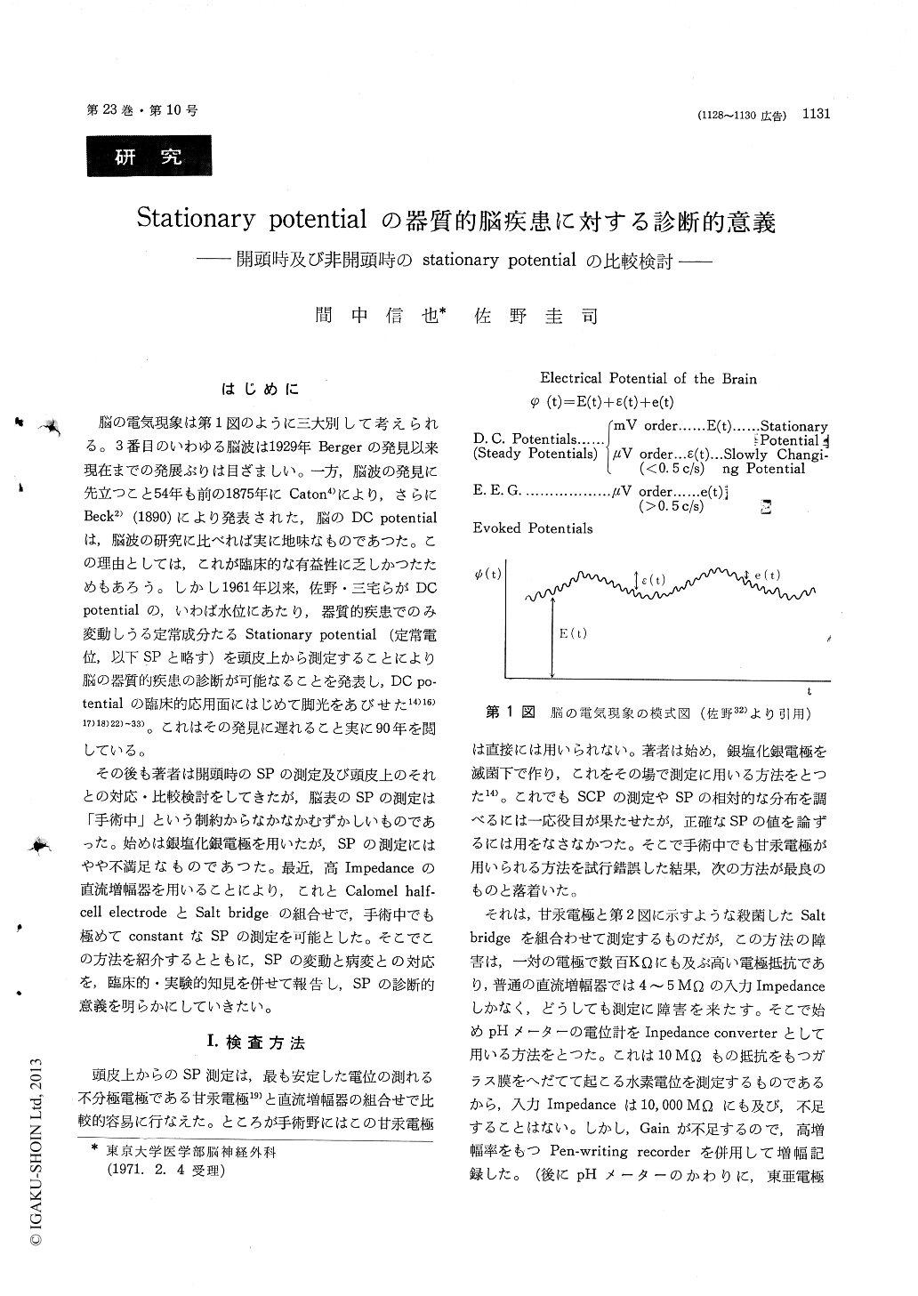
脳の電気現象は第1図のように三大別して考えられる。3番目のいわゆる脳波は1929年Bergerの発見以来現在までの発展ぶりは目ざましい。一方,脳波の発見に先立つこと54年も前の1875年にCaton4)により,さらにBeck2)(1890)により発表された,脳のDC potentialは,脳波の研究に比べれば実に地味なものであつた。この理由としては,これが臨床的な有益性に乏しかつたためもあろう。しかし1961年以来,佐野・三宅らがDCpotentialの,いわば水位にあたり,器質的疾患でのみ変動しうる定常成分たるStationary potential (定常電位,以下SPと略す)を頭皮上から測定することにより脳の器質的疾患の診断が可能なることを発表し,DC po—tentialの臨床的応用面にはじめて脚光をあびせた14)16)17)18)22)〜33)。これはその発見に遅れること実に90年を閲している。
その後も著者は開頭時のSPの測定及び頭皮上のそれとの対応・比較検討をしてきたが,脳表のSPの測定は「手術中」という制約からなかなかむずかしいものであった。始めは銀塩化銀電極を用いたが,SPの測定にはやや不満足なものであつた。最近,高Impedanceの直流増幅器を用いることにより,これとCalomel half—cell electrodeとSalt bridgeの組合せで,手術中でも極めてconstantなSPの測定を可能とした。そこでこの方法を紹介するとともに,SPの変動と病変との対応を,臨床的・実験的知見を併せて報告し,SPの診断的意義を明らかにしていきたい。
The electrical phenomena of the brain can be devided into three categories as illustrated in Fig. 1. The DC potential may be further classified into slowly changing potential (SCP) and stationary potential (SP). The last one is very stable except organic lesion or fetal metablic dis-turbance of the brain or epileptic seizure. Con-versely, the organic cerebral disease could be di-agnosed by recording the SP change. Its possibility was reported by authers already. Subsequently, we tried to record the intracranial SP change insterile condition to correlate with SP on the scalp. In recent years, we could attain to record constant and accurate intracranial SP, using the conbination of calomel half-cell electrodes, sterile salt bridge, high impdance DC amplifires (input impedance is ten thousands MΩ or more) and pen-writing re-corder (see Fig. 2). In conclusion, the distribution and grade of intracranial SP change shows almost identical disposition compairing with the SP change on the scalp. Representive case of meningioma and subcortical tumor are illustrated in Fig. 3 and 5. This fact assures to make diagnosis of intracranial organic disease from recording SP change on the scalp.
Some specific inclination of SP change differed from type of lesion as illustrated in Tab. 1 was dis-covered as the result of recording both intracranial and scalp SP; namely, cortical compression without its damege indicates positive change, cortical com-pression with its damage and/or cortical lesion it-self makes negative change, subcortical and/or deep seated space occupying lesions show positive change, and extensive brain lesion exhibits negative change, respectively. To make clarify this inclination, ex-perimental cortical compression by balloon, cortical compression by balloon, cortical damege and sub-cortical lesion in cat or mongrel dog were investi-gated, and its result is illustrated in Fig. 7 ; in short same tendency that was obtained clinical study was verified. The shema of SP changes by the type of lesions are illustrated in Fig. 8.
Authers proposed resting membrane theory to interpret the origine of stationaly potential (Fig. 9). On the standpoint of view, various SP changes are explainable as illustration of Fig. 10. The crateri-form potential change that is typical in convexity or parasagital meningioma is also introduced finely by this theory.
Recording of SP on scalp is very valuable to predict the nature and depth or size of lesion from the distribution and disposition of SP change. Authers are even of opinion that the diagnostic value of SP recording in intracranial disease excels the EEG.

Copyright © 1971, Igaku-Shoin Ltd. All rights reserved.


