Japanese
English
- 有料閲覧
- Abstract 文献概要
- 1ページ目 Look Inside
はじめに
最近,われわれは外傷性急性硬膜下血腫のうちで種々の事情により手術できなかつたために非観血的に治療をこころみ,頸動脈撮影,脳波所見などで経過を追跡したところ,自然に血腫が消退したと考えられる症例をあいついで経験したので報告する。あわせて外傷性急性硬膜下血腫をふくめた重症脳外傷の病態生理について若干の考察をくわえた。
いわゆる慢性硬膜下血腫や受傷後ある程度の期間をへて,たまたま発見された硬膜下血腫が非観血的に治癒あるいは自然に消退するという報告はそれほど新しいものではなく,文献上でも最近はしばしば散見される1)〜5)。しかし外傷性急性硬膜下血腫において受傷直後より臨床神経学的所見脳波所見,頸動脈撮影にてその経過を追跡しえた例はほとんどない。慢性硬膜下血豚の自然治癒,あるいは非観血的消退はその発生機序とともに興味ある問題である。しかし一方,急性硬膜下血腫の自然治癒の事実は重症脳外傷の治療についていくつかの重要な問題を提起していると考えられる。
Four cases of traumatic acute subdural hematoma which had spontaneous resolution were presented.
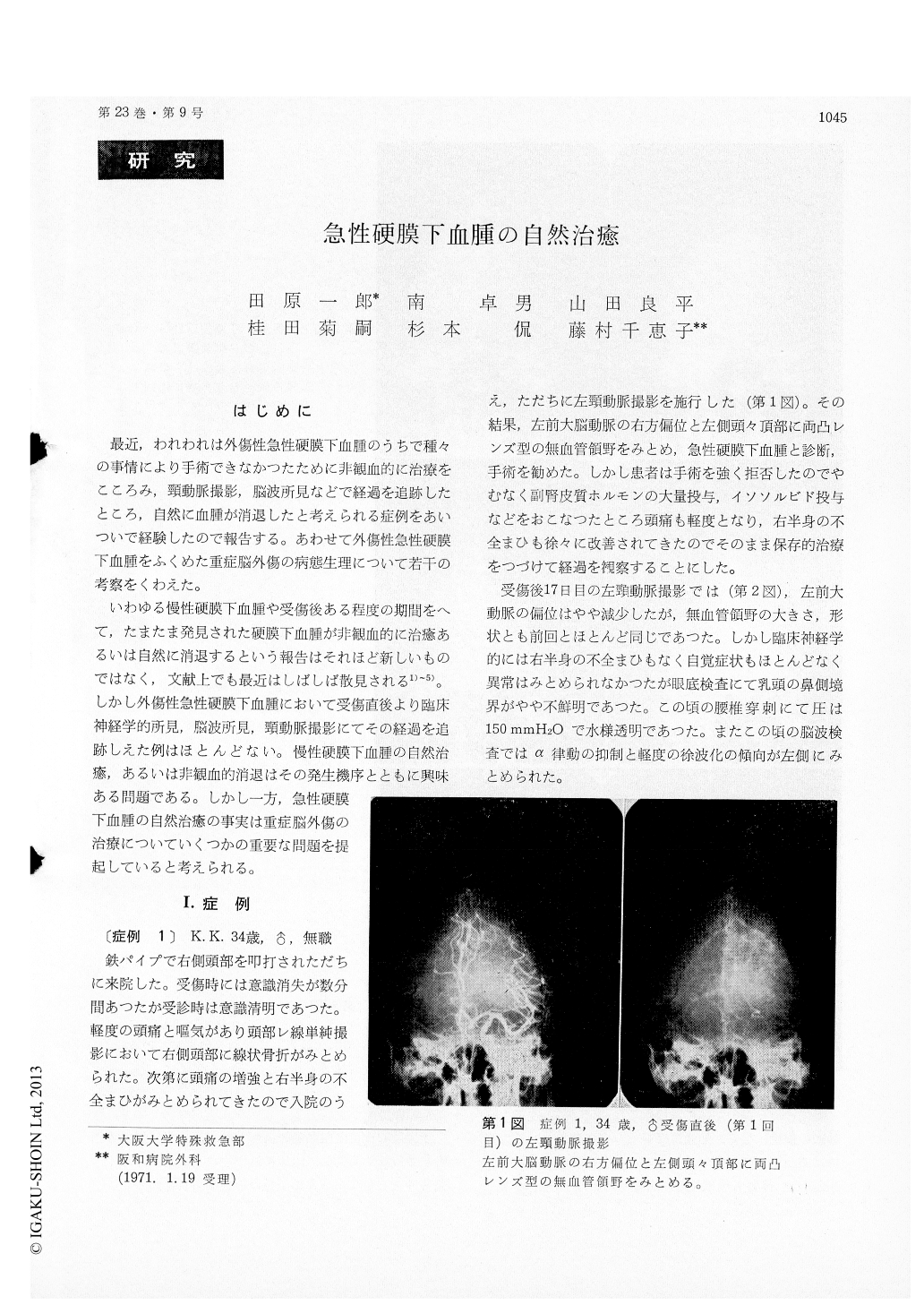
Case 1, a 34-year-old man, struck his head a few hours prior to admission, when he had headache, nausea, and incompleter hemiplegia. Carotid angio-graphy was performed immediately, which re-vealed an avascular area of biconvex type in the left parietotemporal region. The patient refused the surgery and followed with conservative treat-ment. Though the angiogram of the 17th day after admission revealed the unchanged size of avascular area, the patient's condition markedly improved in the successive days. On the angio-gram of the 53rd and 92nd hospital day, the sub-dural hematoma was gradually decreased in size into convex-concurve type and finally confirmed to have been disappeared. The patient was quite well and later examination of EEG showed no evidence of neurological deficit.
Case 2, a 59-year-old man, struck his head and visited our clinic immediately. On the second posttraumatic day, when he complained of increas-ing headache, carotid angiogram revealed diffuse subdural collection in the right parietal region. Following the supportive therapy in a few suc-cessive days, his complaints were subsided. Angio-gram of the 14th and 42nd day after trauma showed the gradual resolution of the hematoma.
Case 3, a 38-year-old man, automobile accident 5 days before admission, when he had oneset of mild unconciousness and anisocoria. Carotid angio-gram suggested the left-sided hematoma of tem-poral subdural space. It was no longer than 15 days in this case that the hematoma was disappeared angiographically. He had been recovered from the neurological impairment.
Case 4, a 49-year-old man, struck his head and visited our clinic immediately. He complained of headache and the slight depression of the level of consciousness was observed. Then carotid angio-graphy was performed, which revealed an avascular area of convex-concurve type and the displacement of the anterior cerebral artery completely fainted. The later examination and EEG showed no evid-ence of neurological deficit.
Further consideration were added about the patient with acute subdural hematoma who were treated by surgical intervention. And the possible relation-ship between pathology of subdural hematoma and medical treatment was discussed.
The present experiences suggest that, at first,compression effect to brain by subdural mass is not the major factor contributing to neurological deficit or brain stem function. Sustained or chronic sub-dural hematoma has been occasionally disclosed with-out neurological disorder, that is true about the acute hematoma as presented here. At the second, it is suggested that acute subdural hematoma can be unexceptionally dissolved in a few months with-out any sequel. It is not the aim of this paper to claim that supportive therapy will replace opera-tion in every case of acute subdural hematoma. We urge that the surgical intervention is not an only potential treatment upon the pathology of acute subdural hematoma.
Copyright © 1971, Igaku-Shoin Ltd. All rights reserved.