Japanese
English
- 有料閲覧
- Abstract 文献概要
- 1ページ目 Look Inside
はじめに
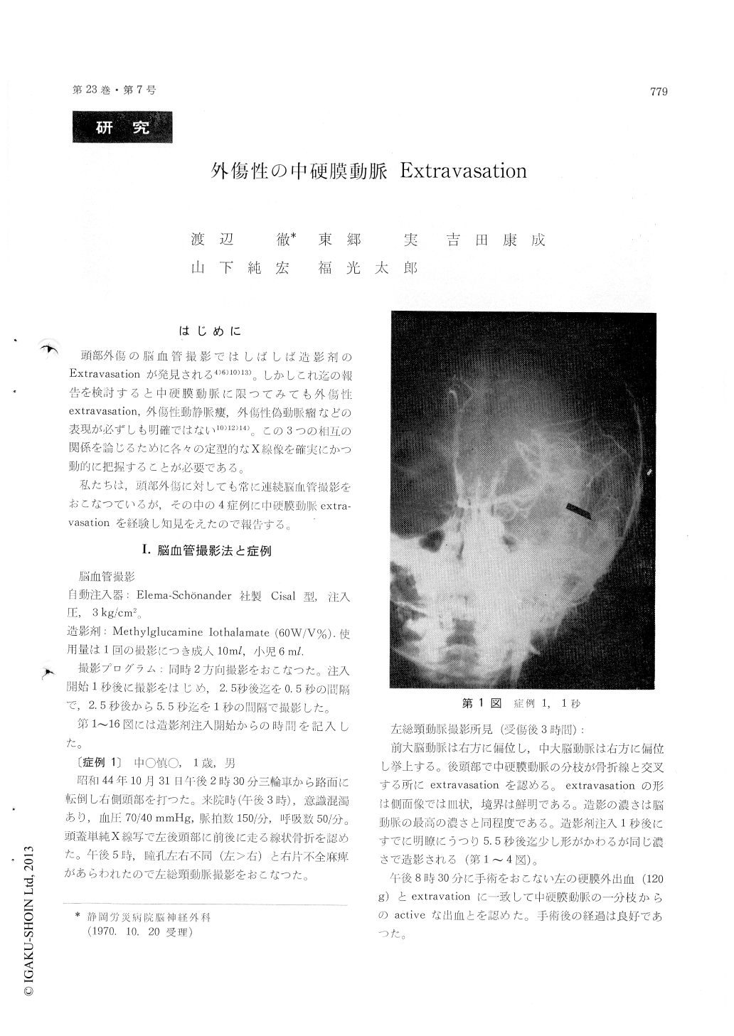
頭部外傷の脳血管撮影ではしばしば造影剤のExtravasationが発見される4)6)10)13)。しかしこれ迄の報告を検討すると中硬膜動脈に限つてみても外傷性extravasation,外傷性動静脈瘻,外傷性偽動脈瘤などの表現が必ずしも明確ではない10)12)14)。この3つの相互の関係を論じるために各々の定型的なX線像を確実にかつ動的に把握することが必要である。
私たちは,頭部外傷に対しても常に連続脳血管撮影をおこなつているが,その中の4症例に中硬膜動脈extra—vasationを経験し知見をえたので報告する。
Four cases of traumatic extravasation of contrast medium from the middle meningeal artery were studied in serial angiography.
Common carotid angiography was done within six hours after head injury. Extravasations were found where the middle meningeal artery crossed the linear fracture of skull.
They were visible within 2 seconds following initial injection of contrast medium. The shape, size, and opacity were not uniform. The posterior border of the extravasated contrast medium was sharply defined in lateral view. The opacity, not less than that of cerebral vessel in the arterial phase, was constant for more than 3.5 seconds, accom-panied with some change in shape and size.
At operation, the extravasation in two cases was confirmed as an active bleeding from the injured meningeal artery without any evidence of false aneurysm formation or arteriovenous fistula.

Copyright © 1971, Igaku-Shoin Ltd. All rights reserved.


