Japanese
English
- 有料閲覧
- Abstract 文献概要
- 1ページ目 Look Inside
はじめに
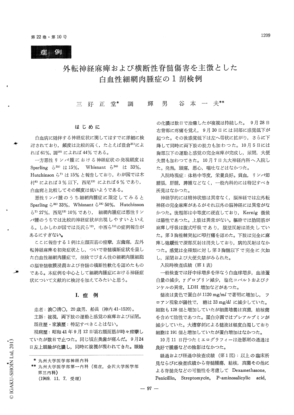
白血病に随伴する神経症状に関してはすでに詳細に検討されており,頻度は比較的高く,たとえば豊倉21)によれば61%,調18)によれば44%である。
一方悪性リンパ腫における神経症状の発現頻度はSparlingら20)は15%, Whisnantら24)は33%, Hutchinsonら7)は15%と報告しており,わが国では木村8)によれば3%以下,西尾13)によれば6%であり,白血病と比較してその頻度は低いようである。
A 20-year-old Japanese man, admitted to Depart-ment of Neurology of Kyushu University Hospital on October 7, 1966, had been well until one month prior to admission, when he noticed spasm of left facial muscles for several days. On September 25, he became to have furunkle of left eye lid and double vision. On September 30, back pain appeared at the level of Th-3, and within a few days sensory loss below the level of Th-3 and paraplegia devel-oped.
Examination on admission revealed left abducens nerve palsy, paraplegia, sensory loss below the level of Th-3 and double incontinences. Lymphadeno-pathy, hepatomegaly and splenomegaly were not detected. Lumbar cerebrospinal fluid showed marked increase of protein with Froin's sign, pleocytosis and decrease in sugar centent, but the CSF in cisterna magna disclosed only pleocytosis without increase in protein content. Myelography was performed, but failed to disclose the blocking. On November 13, he had acute melena resulting in marked anemia. Intermural tumors in stomach were seen by gastro-intestinal fluoroscopy. Hepatomegaly, hemorrhagic diathesis and emaciation appeared. Abnormal cells ranging from 5 to 10 per cent of the whole leuk-ocytes were found in the peripheral blood smear. These abnormal cells were large and irregular in shape, with vacuoles in abundant cytoplama. The nuclei were ovoid or irregularly round in shape with nuclear chromatin showing fine network as well as a few large nucleolus. He was diagnosed as leukemic reticulosarcomatosis based on these cells.
On December 20, dysphagia was noted, followed by right facial nerve palsy in two days. He died on December 25, 1966.
Autopy examination showed that liver was mark-edly enlarged with infiltraion of reticulosarcomatous cells. Other organs, spleen, kidneys, pancreas, suprarenal glands, bone marrow, meninges, sto-mach, small and large intestine, testis and postperi-toneal and inguinal lymph nodes were also infiltrated by the same cells. While there was a softening and necrosis in spinal cord from segment Th-3 to Th-8, no tumor was seen in epidural space. How-ever, spinal and cranial nerves and leptomeninges of brain and spinal cord were diffusely infiltrated by the reticulosarcomatous cells.

Copyright © 1970, Igaku-Shoin Ltd. All rights reserved.


