Japanese
English
- 有料閲覧
- Abstract 文献概要
- 1ページ目 Look Inside
I.はじめに
前報3)ではアンマ針を電極として用い,定速度電極刺入法により測定したネコの脳の部位の差によるimpe—danceの変動につき報告した。
最近定位脳手術時,その破壊部位決定にimpedance法を利用1),あるいは脳腫瘍の性状の相違によるimpedanceの値の差の検出の試み2)など,脳神経外科臨床領域においても,脳の電気impedanceに関する研究発表が相次ぎ,ながらくの間あまり顧りみられなかつた本分野の研究が,再び世の注目を浴びるに至つた感があるが,われわれも前報以降,動物実験と並行して人脳impedanceの測定を行ない,またこの目的のための増幅器および測定用特殊電極を作製した。
This is the second report mainly concerned with changes in the brain tissue impedance measured both in animals and in patients with centrally located malignant tumor (s). Impedance curve obtained from a Parkinsonian patient under-going stereotaxic sur-gery was also reported.
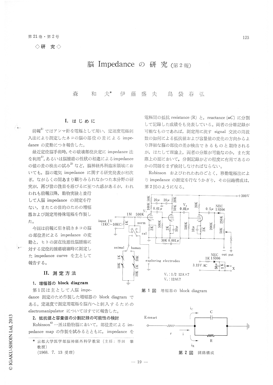
For this purpose, specially designed amplifier was used (Fig. 1). Impedance was measured by means of a roving electrode and with the sine wave cur-rent of 10 kc.
Table 1 summarizes impedance values obtained from the various thalamic nuclei in 16 cats. In this table, impedance value of cortex was taken as a con-trol and expressed as 100. Thalamic values were cal-culated in terms of percentage for the control. The maximum difference in impedance in these thalamic nuclei was within the range of 500Ω-600Ω, and the value of the impedance recording has not yet proved to be a useful method in differentiating and locating these thalamic nuclei. However, by using the impedance technique. we could differentiate the thalamic gray matter from the surrounding struc-tures with accuracy. While the measuring electrode was being introduced into thalamus, the impedance showed a marked and characteristic decrease and after traversing the thalamus, the impedance started rising again. Consequently, the curve made by measuring the tissue impedance continuously from the cortex to deep in the subthalamic region, exhibited in the shape of "a frying pan" while the electrode was passing the thalamus (Fig. 3 and Fig. 4).
In 3 cases with centrally located malignant tumor (s), a probe for radiofrequency coagulation was served as the measuring electrode and impedance was being recorded during the introduction of the probe into tumor (s) (Fig. 5 and Fig. 6). In this case, the impedance changes were indistinct as compared with those obtained in animal experiments, presumably because we used the big probe as the active electrode. However, the location of corpus callosum and lateral ventricle were well identified in the impedance trace. Moreover, there was a marked increase in impedance in the thalamic region which might reflect the cer-ebral pathology and electrocoagulation was carried out at the sites where impedance showed such ab-normal high value.
Fig. 7 showes impedance curve obtained during the stereotaxic thalamotomy for Parkinsonism. In this case, a small (0.5 mm in diameter) insulated stainless wire was used as the active (measuring) electrode and impedance changes were recorded bipolarly between this active electrode and the coagulation probe. The myelin-rich tissues, the in-ternal capsle (CI) and the Forel H field and so on, reflected as high value in impedance which gave us the sharp landmarks to deciding the target during the stereotaxic surgery.
It was concluded that the use of impedance mea-surements during stereotaxic surgery is of potential value to identify the cerebral structures and to make a lesion with safe and more accuracy.
This study was supported in part by NIH Grant No. 06553.
Copyright © 1969, Igaku-Shoin Ltd. All rights reserved.