Japanese
English
- 有料閲覧
- Abstract 文献概要
- 1ページ目 Look Inside
I.緒言
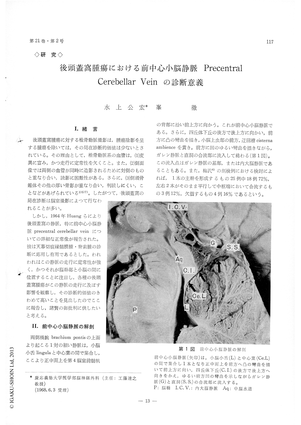
後頭蓋窩腫瘍に対する椎骨動脈撮影は,腫瘍陰影を呈する腫瘍を除いては,その局在診断的価値は少ないとされている。その理由として,椎骨動脈系の血管は,(1)変異に富み,かつ走行に定常性を欠くこと。また,(2)側面像では両側の血管が同時に造影されるために対側のものと重なり合い,読影に困難性がある。さらに,(3)側頭骨錐体その他の厚い骨影が重なり合い,判読しにくい,ことなどがあげられている1)5)7)。したがつて,後頭蓋窩の局在診断は脳室撮影によつて行なわれることが多い。
しかし,1964年Huangらにより後頭蓋窩の静脈,特に前中心小脳静脈precentral cerebellar veinについての詳細な正常像が報告された。彼は天幕切痕縁髄膜腫・脊索腫の診断に応用し有用であるとした。われわれはこの静脈の走行に定常性が強く,かつそれが脳幹部と小脳の間に位置することに注目し,各種の後頭蓋窩腫瘍がこの静脈の走行に及ぼす影響を観察し,その診断的価値のきわめて高いことを見出したのでここに報告し,諸賢の御批判に供したいと考える。
1) The precentral cerebellar vein begins between the lingula and the central lobe and runs upwards behind the anterior medullary velum in midline.
2) The normal course and configuration of this vein was described. The minimal distance from this vein to the posterior aspect of the clivus 38 mm with a range of 31 to 43 mm measured in 40 normal cases.
3) The deformities and displacement of this vein indicate the changes in the aqueduct and the upper part of the fourth ventricle caused by tumors of mesencephalon, pons and cerebellum.
4) Diagnostic value of this vein is highly evalu-ated in the vertebral angiography by the authors.

Copyright © 1969, Igaku-Shoin Ltd. All rights reserved.


