Japanese
English
- 有料閲覧
- Abstract 文献概要
- 1ページ目 Look Inside
I.はじめに
頭蓋内hemangioblastomaは比較的稀れなもので,全脳腫瘍の約1-2%の割にみられる。しかも,その大部分は小脳に生じ,時に網膜の血管腫の他,肺,膵,肝,腎にも血管腫や嚢胞を伴い,あるいはまた,polycytemiaを伴いLindau v. Hippel’s diseaseとしてしられている。このhemangioblastomaがテント上に生ずることは,はなはだ稀れなこととされ,最近のMorello1)らの報告によれば,今日までに報告されているものは全部で6例に過ぎないという。東大脳神経外科の2,400余例の脳腫瘍のうち,hemangioblastomaと診断されたものは27例で,そのなかの2例がテント上に生じたもので,それぞれ前頭葉,頭頂後頭葉にみられた。これらテント上hemangioblastoma 2例について,臨床所見,手術所見および病理組織所見を述べ,文献的考察を行ないたいと思う。
Supratentorial hemangioblastomas are encountered very rarely. However, out of the 27 cases of in-tracranial hemangioblastomas admitted at the Dept. of Neurosurgery, University of Tokyo Hospital, two cases turned out to be supratentorial hemangio-blastomas and their locations were left frontal lobe and the left parieto-occipital lobe respectively. These two cases were presented in this report and the authors have described and discussed on some clinical aspects of the supratentorial hemangioblastomas.
A short history of the cases are as follows:
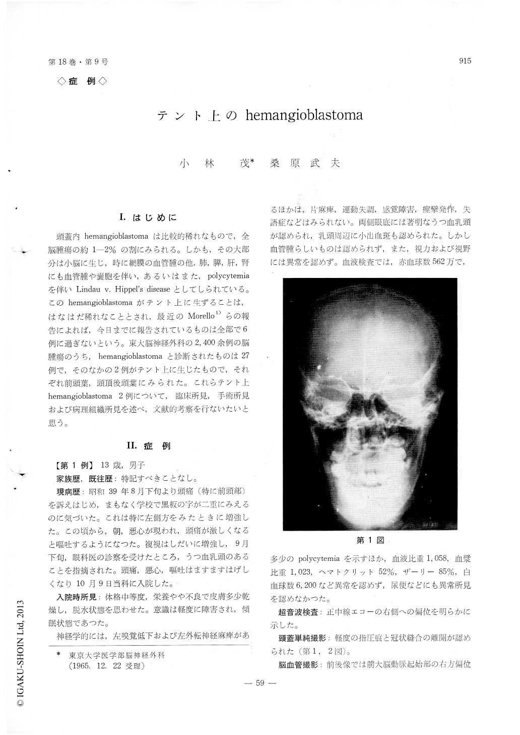
Case 1. 13 years old boy, with complaints of headache, nausea, vomiting, diplopia and decreased visual acuity for the duration of one month, was admitted. At the time of admission, objective find-ings were left abducens palsy, left hyposmia, bila-teral choked disks and in a state of lethargy.
During the operation, a hen's egg sized cystic tumor locating in the postero-lateral part of the left frontal lobe was found. Evacuation of the cyst and removal of the mural nodule was done. Histolo-gical examination proved to be a typical hemangio-blastoma.
Post-operative course was uneventful and all the complaints have disappeared at the time of discharge.
Case 2. 31 years old man. Prior to two years of his admission, fine movements of his right hand became gradually unskillful followed by gradual manifestations of decreased visual acuity, headache, disturbance of memory and speech disturbances.
At the time of admission, objective findings were increased intracranial pressure, right homonymous hemianopsia and Gerstmann's syndrome.
During the operation, a cystic tumor locating in the parieto-occipital lobe was found subcortically. It was an enormous cyst, about a fist-sized, brownish-red in color, spongy in consistency and mural nodule was located on a part of the cystic wall. The cyst contents were evacuated and the nodule was partially removed.
Post-operative course was uneventful and the patient was discharged from the hospital on the 21 st post-operative day with hemianopsia persisting.
Copyright © 1966, Igaku-Shoin Ltd. All rights reserved.