Japanese
English
- 有料閲覧
- Abstract 文献概要
- 1ページ目 Look Inside
I.はじめに
脳の血管芽腫hemangioblastoma (テント上,後頭蓋窩のものを含む)は,脳腫瘍のなかでの頻度は比較的低く,われわれの教室の2,500例の脳腫瘍のうち,30例で,1.2%にすぎない。しかし,この腫瘍は,他の脳腫瘍とは異なつた,種々の興味ある特徴があり,脳腫瘍のなかでも特異な存在である。そこで,われわれの教室の30例の,脳のhemangioblastomaについて総括し,特に興味ある点について考察を加えたい。
本論にはいる前に,「Lindau病」あるいは,「Lindau腫瘍」という名称について,まず,われわれの見解を述べておきたい。これについては,べつの機会にすでにくわしく論じた34)ので,ここでは要点のみにとどめるが,臨床例については「Lindau病」という名称は適当でなく,Lindauの報告13)が,剖検例についてのものであるから,病理学的な概念にとどめるのがもつとも妥当であると考えられる。もし「Lindau病」という名称を,臨床的に用いる場合は,angiomatosis retinae (von Hippel病)を伴つた,脳や脊髄のheman—gioblastomaに限定すべきものと考えている。脳,脊髄のhemangioblastomaやvon Hippel病は,むしろ広く「中枢伸経系のhemangioblastoma」という範疇にいれて考えるのが,Lindauの意図をくんだ,もつとも適切なものであろう。
The authors reported 30 cases of hemangioblstomas of the brain which they have experienced. The cases were found more frequently in males, the male-to-female ratio being 2: 1.
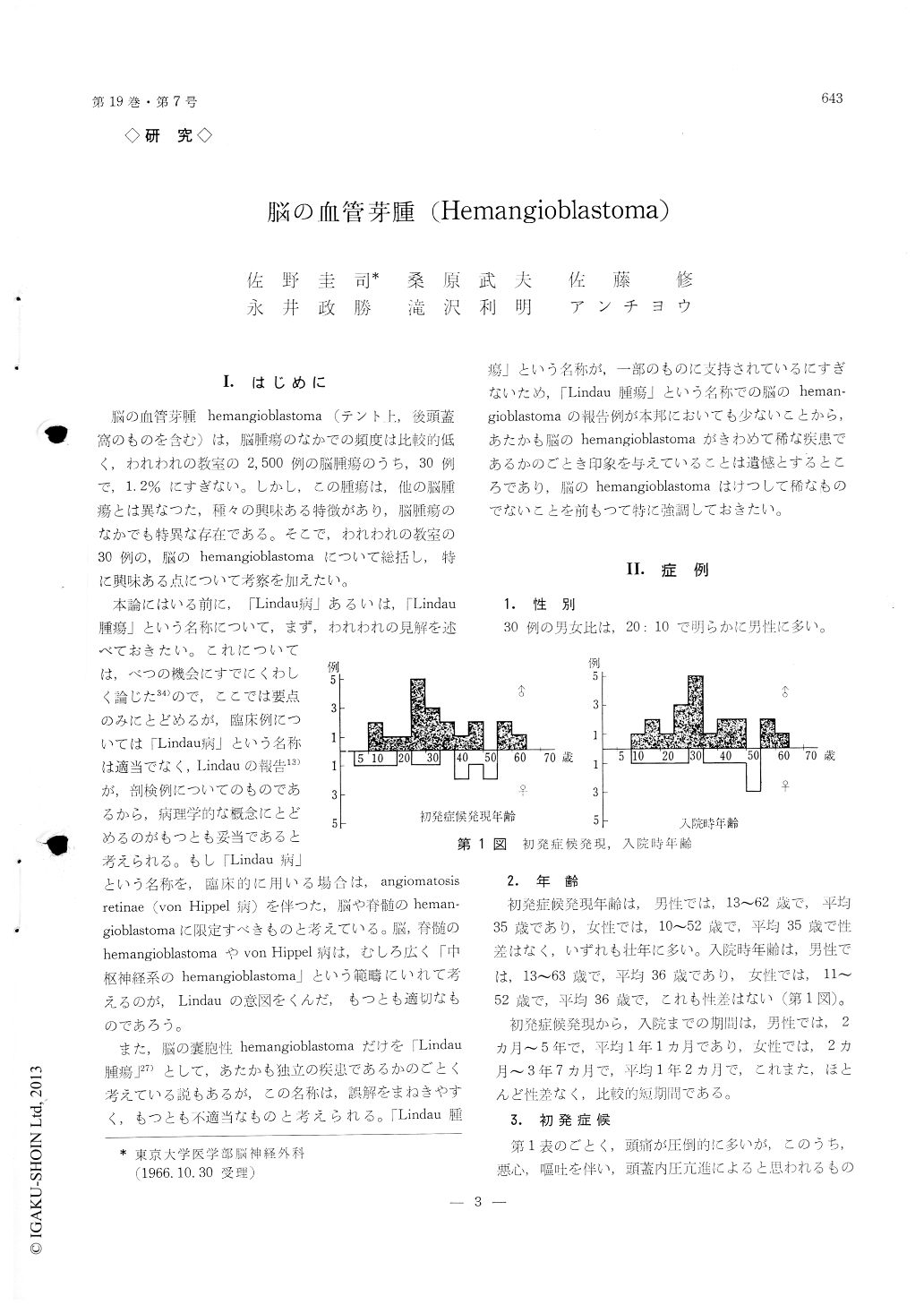
The mean age at the onset of the symptoms and signs was 35 years of age and the duration of the symptoms and signs before admission was about one year.
Among the initial symptoms and signs, headache was most frequent, which, however, was not supposed due to be the increased intracranial pressure. Headache was also most frequently found on admission,which was definitely caused by the increased intracranial pressure. In the cases of hemangioblastomas in the posterior fossa, the main neurological findings on admission were those of the cerebellum or the brain stem, which made the diagnosis of tumor in the poste-rior fossa easy. In order to diagnose the nature of the tumor accurately, the vertebral angiography was man-datory. The authors classified the vertebral angiograms of the tumors in the posterior fossa into three groups ; namely, (A) one with a solitary shadow of the tumor, (B) one with a diffuse shadow of the tumor and (C) one without shadow of the tumor, showing only dis-location of the vessels.
Among 30 cases, 2 cases showed angiomatosis retinae (von Hippel's disease) and one case was found to have an angioma of the chorioidea in the carotid angiogram.
Familial occurrence was noted in 3 cases of the two familial pedigrees. One case had cystic tumors of the bilateral kidneys with hypertension. Eryth-rocytosis was noted in 10 cases (33. 3%), being more frequently found in solid tumors than in cystic tumors (40: 26. 7%). In 8 cases with erythrocytosis, the follow-up studies were done. The results were as follows: In 3 cases of total extirpation of the tumor, erythrocytosis disappeared after the operation. Among 5 cases of partial exision and/or decompression with postoperative irradiation, 3 cases showed disappearance of erythrocytosis after the treatment and 2 cases showed no significant change in the blood picture.
Hemangioblastomas were most frequently found in the cerebellum (71%), then in the area postrema (13%), and rarely in the cerebral hemisphere (6.5%) and the medulla oblongata (6.5%).
Total extirpation of the tumor was always the treatment of choice, whenever feasible. When this was unable and only partial exision or decompression was able to by performed, postoperative irradiation seemed remarkable effective in improving the sym-ptoms and signs and in preventing recurrence.

Copyright © 1967, Igaku-Shoin Ltd. All rights reserved.


