Japanese
English
- 有料閲覧
- Abstract 文献概要
- 1ページ目 Look Inside
I.はじめに
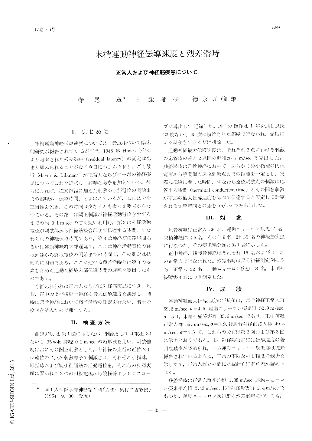
末梢運動神経伝導速度については,最近相ついで臨床的研究が報告されているが1)〜4),1948年Hodesら5)により考案された残差潜時(residual latency)の測定はあまり顧みられることがなく今日におよんでおり,ごく最近Mavor & Libman2)が正常人ならびに一部の神経疾患についてこれを追試し,詳細な考察を加えている。彼らによれば,従来神経に加えた刺激から筋電位の開始までの潜時が「伝導時間」とよばれているが,これはやや正当性を欠き,この時間は少なくとも次の3要素からなつている。その第1は閾上刺激が神経活動電位を生ずるまでの約0.1msecのごく短い利用時,第2は神経活動電位が刺激部から神経筋接合部まで伝達する時間,すなわち真の神経伝導時間であり,第3は神経筋伝達時間あるいは運動神経終末部遅延で,これは神経活動電位の終板到達から終板電位の開始までの時間で,その測定は技術的に困難である。ここに述べる残差潜時とは第3の要素を含めた運動神経終末部伝導時間の遅延を算出したものである。
今回われわれは正常人ならびに神経筋疾患につき,尺骨,正中および後脛骨神経の最大伝導速度を測定し,同時に尺骨神経において残差潜時の測定を行ない,若干の検討を試みたので報告する。
We measured the maximum conduction velocities of ulnar nerve, median nerve and posterior tibial nerve in the normal and the neuromuscular diseases and estimated the residual latency of ulnar nerve. In the normal group, mean maximum conduction velocity of ulnar nerve was 59.6±4.5 m/sec, whereas the mean velocity in the neuromuscular disease was 52.9±5.1m /sec, which was slower than in the normal. In contrast with these two groups the velocity in the group of peripheral nerve disturbances markedly decreased to 35.6m/sec. The normal residual latency was 1.38m/sec but it slightly delayed in the neuromuscular diseases and showed 2.43msec.
We also discussed the relation between the conduction velocities and the pathological changes of peripheral nerve in two clinical cases of spinal progressive muscle atrophy.

Copyright © 1965, Igaku-Shoin Ltd. All rights reserved.


