Japanese
English
- 有料閲覧
- Abstract 文献概要
- 1ページ目 Look Inside
II.絶食による変化
I.緒言
精神身体症の病態論追及の一部として,先に向精神薬による海馬体神経細胞の変化を観察し,糸粒体,endoplasmic reticulum,核膜等に変化の起こることを報告した。しかし,臨床的には今までの向精神薬は精神身体症の治療に対しては効果が不良であり,経験的に絶食が有効であることがわかつている。そこで,著者は絶食(飢餓)時に海馬体神経細胞がいかなる変化を示すかについて電子顕微鏡により観察した。
As a link in the series of studies in pursu-ance of the pathology of psychosomatic dis-ease, we have studied the changes in the hip-pocampal nerve cells after administration of psychotropic agents and reported that chan-ges were brought about in the mitochondria, the endoplasmic reticula and the nuclear membrane of such cells. But clinically, the psychotropic remedies in current use are known to be not so effective as disired and rather simple fasting is found more efficaci-ous for treating such diseases. Therefore, we undertook a study of the hippocampal cells of male mature rats on the 3 rd day of fast-ing and the 2 nd and the 7 th days after breaking the fast, under an electron micro-scope, with the aim of ascertaining the effect of fasting upon the cells.
The result were in summary as follows;
A. Changes in the clear nerve cells.
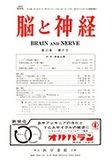
1) The fine granules in the cytoplasm were found decreased during fasting and were still subnormal in number on the 2 nd day after it, but reincreased to the normal by the 7 th day.
2) Undersized and pycnotized mitochondria were observed during fasting, but on the 7 th day after it, the mitochondria were not percep-tibly different from the normal.
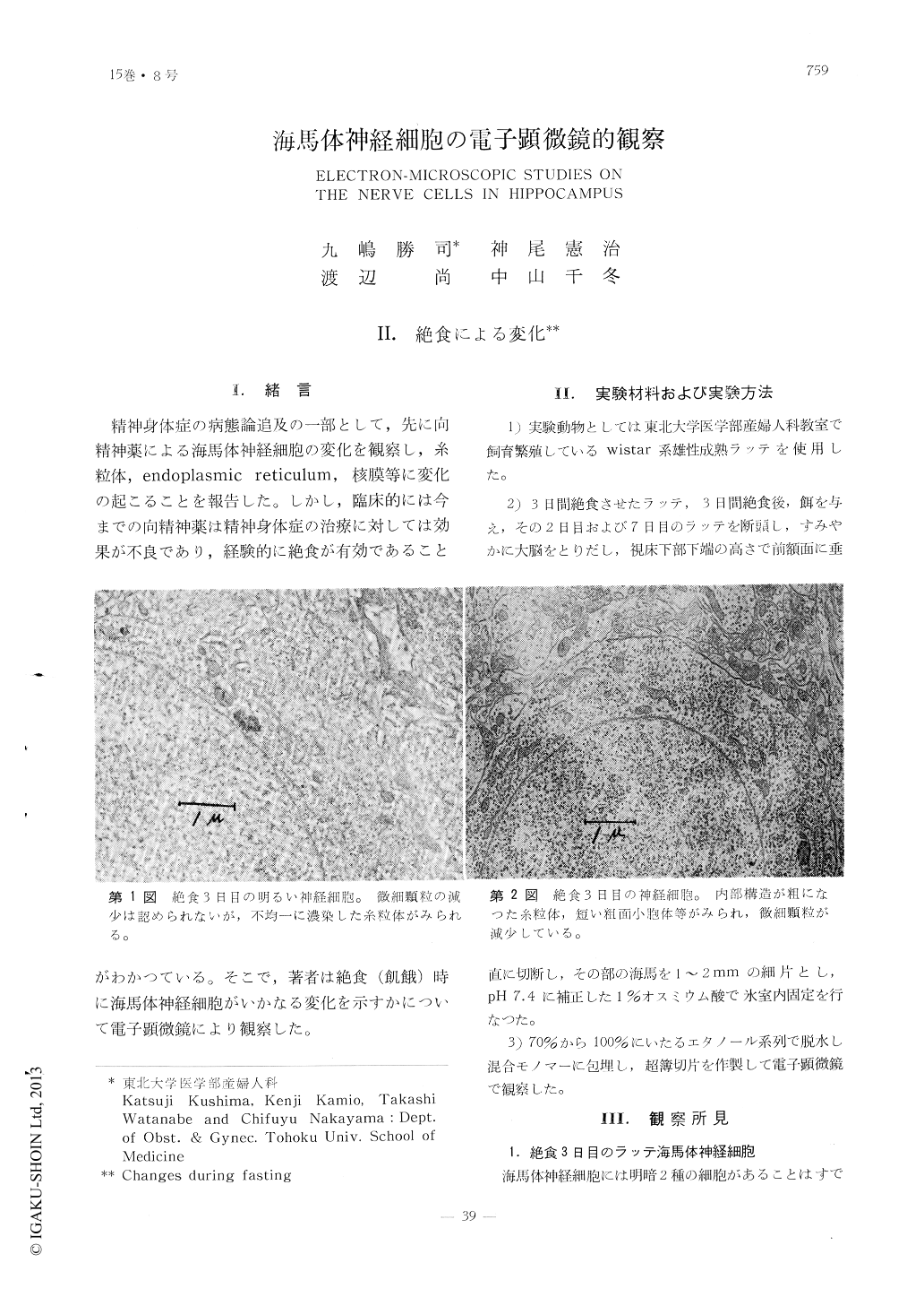
3) Some of the endoplasmic reticula were shortend and the fine granules on the surface of the roughsurfaced ones were decreased during fasting, but on the 7 th day after it, they were very nearly normal.
4) The nuclear membrane was not percepti-bly changed during fasting but after break-ing the fast, it was often irregularly uneven. B. Changes in the dark cells.
Morphologically, these cells showed no per-ceptible change during fasting. On the 2 nd day after it, the endoplasmic reticula were increased in lamellar arrangement, but the decrease of fine granules on them was not clearly observable.
Copyright © 1963, Igaku-Shoin Ltd. All rights reserved.