Japanese
English
- 有料閲覧
- Abstract 文献概要
- 1ページ目 Look Inside
まえがき
上顎癌が上のほうに進んで眼球突出や視力障害を生じたり,頭蓋底を破壊しさらに頭蓋内に侵入することがある。癌が頭蓋底をおかすさい,その部位を通過している脳神経を傷害するため脳神経症状や,時には複雑な多発性脳神経麻痺症候群を惹起する。
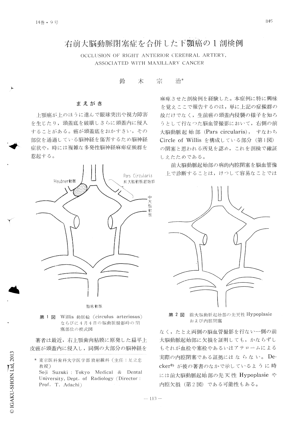
著者は最近,右上顎歯肉粘膜に原発した扁平上皮癌が頭蓋内に侵入し,同側の大部分の脳神経を麻痺させた剖検例を経験した。本症例に特に興味を覚えここで報告するのは,単に上記の症候群の故だけでなく,生前癌の頭蓋内侵襲の様子を知ろうとして行なつた脳血管撮影において,右側の前大脳動脈起始部(Pars circularis),すなわちCircle of Willisを構成している部分(第1図)の閉塞と思われる所見を認め,これを剖検で確証しえたためである。
This 35-year-old female began to complain of pain in the secound molar region of right maxilla in January of 1959. Extraction of the secound molar tooth resulted in a persis-tent ulcer with a deep fistula in the socket of the extracted tooth. The fistula was soon perforated into the right antrum and a flat tumor arised from the margin of the ulcer.
She was treated by implantation of radon seeds (12mc) and X-irradiation (3400 r thr-ough two fields) with subsequent disappea-rance of the tumor and pain.
On August 16, 1960 she again presented herself to the Radiological Clinic with pain in the cheek on right side and marked trismus. The inspection disclosed a large recurrent tumor on the palate and gingiva. The neurological examination at that time was negative. On November 14, 1963 implantation of radon seeas (17mc) into the tumor wascarried out.
In beginning of December of 1960 the right eye became blind and paralysed. and loss of sensation in the first division of right trigemi-nal nerve manifested itself. Lumbar puncture, however, was essentially negative.
The neurological examination in March of 1961 revealed unilateral paralysis of eight cranial nerves (I~VIII) on right side.
Right carotid angiography on April 4, 1961 disclosed an occlusion of the right anterior cerebral artery in Pars Circularis. The anterior communicating artery and both anterior cerebral arteries were visualised on left carotid angiogram. The site of the occlusion should be distal to the origin of Heubner's artery and proximal to the anterior communicating artery, because the right Heubner's artery was demonstrated on the right carotid angiogram. The above-mentioned anatomical location of the occlusion mightexplain the reason why the patient had de-veloped no sign of the circulatory disturbance of the right cerebral hemisphere.
In late April of 1961 she developed paralysis of the left lower extremity and profound mental disorder, and became comatose on May 4, 1961 and expired on the following day.
Autopsy disclosed that the tumor destroyed the base of the skull on right side and invaded into the cranial fossae. The tumor mass in the right middle cranial fossa pushed and displaced the temporal lobe of the right cerebral hemisphere upwards. The lumen of the right anterior cerebral artery was completely occluded by thrombus from its origin to the periphery. The lumen of the anterior communicating artery and left anterior cerebral artery was patent and normal.

Copyright © 1962, Igaku-Shoin Ltd. All rights reserved.


