Japanese
English
- 有料閲覧
- Abstract 文献概要
- 1ページ目 Look Inside
緒言
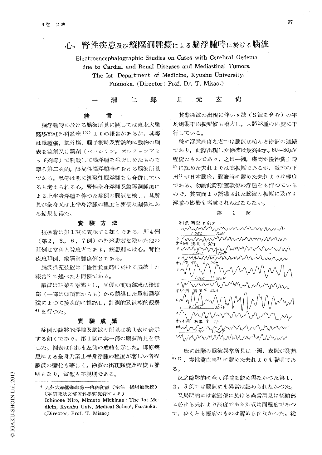
腦浮腫時に於ける腦波所見に關しては東北大學醫學部桂外科教室1)2)よりの報告があるが,其等は腦腫瘍,腦外傷,腦手術時及實驗的に動物の腦表を空氣又は藥剤(ペニシリン,ズルフォンアミッド剤等)で刺戟して腦浮腫を生ぜしめたもので寧ろ第二次的,限局性腦浮腫時における腦波所見である。私等は明に汎發性腦浮腫をも合併していると考えられる心,腎性全身浮腫及縱隔洞腫瘍による上半身浮腫を伴つた症例の腦波を検し,其所見が全身又は上半身浮腫の程度と密接な關係にある結果を得た。
Recording the E.E.G. on cases with cardial and renal diseases (13 cases) and mediastinal tumors (2 cases), we obtained following results showing a close relationship between the ele-ctroencephalographic change and clinical oede-ma due to the above mentioned diseases.
1) In the cases with no clinical oedema, the abnormality in the E.E.G. was not observed.
2) In the cases with clinical oedema, the slow waves with high amplitude and the irregu- larity of wave type appeaed in the frontal, parietal and occipital areas.
With the increasing of clinical oedema. these tendencils became more and more remarkable.
3) These changes in the frontal area were marked than these in the occipital area.
Therefore, in some cases with clinical oedema the mean value of amplitude of the α wave, including the δ wave, in the frontal area became greater than that in the occi- pital area.
4) With the decreasing of clinical oedema, the above mentioned changes were reversible.
5) These changes in the E.E.G. may be observ- ed resulting from cerebral oedema.

Copyright © 1952, Igaku-Shoin Ltd. All rights reserved.


