Japanese
English
- 有料閲覧
- Abstract 文献概要
- 1ページ目 Look Inside
緒言
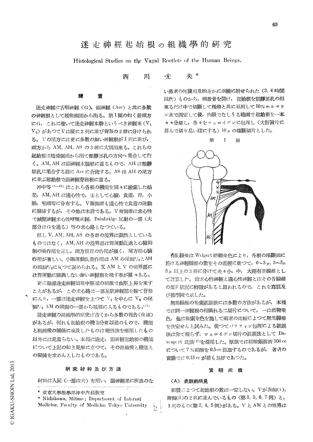
迷走神經は舌咽神經(G),副神經(Acc)と共に多數の神經根として延髓側面から出る。第1圖の如く最頭方にG,これに續いて迷走神經本幹というべき神經束(V1V2)があつてVは縦に2列に並び背腹の2群に分けられる。Vの尾方には更に多數の細い神經根が1列に並び,頭方からAM, AH, ASの3群に大別出來る。これらの起始根は延髓側面から出て頸靜脈孔の方向へ集合して行く。AM, AHは副神經末腦根に當るもので,AHは頸靜脈孔に集合する前にAccに合流する。ASはAHの尾方に並ぶ起始根で副神經脊髓根に當る。
冲中等(1-15)はこれら各根の機能を別々に檢索した結果,AM, AHは遠心性で,主として心臟,食道,胃,小腸,喉頭等に分布する。V腹側群も遠心性で食道の運動に關係するが,その他は未詳である。V背側群は求心性で減壓神經求心性呼吸神經,Bainbridge反射の一部(大部分はGを通る)等の求心路となつている。
但しV, AM, AH, ASの各群の境界は劃然としているものではなく,AM,AHの境界部は胃運動促進と心臟抑制の兩作用を示し,頭方程胃の作用が強く,尾方程心臟作用が著しい。小腸運動促進作用はAMの尾側1/3とAHの頭側2/3に互つて認められる。又AMとVの境界部には胃運動に關與しない細い神經根を殘す事が?々ある。
更に頸部迷走神經切斷中樞端の刺激で血壓上昇を來すことがあるが,この求心路は一部星?神經節を經て脊髓に入り一部は迷走神經を上つてV1を中心にV2の尾側1/3AMの頭側の一部から延髓に入るものである。(12)
迷走神經の組織學的研究は古くから多數の報告(後述)があるが,何れも起始根の機能分離以前のもので,機能と組織像の關係に論及したものは變性法を應用したもの以外には見當らない。本報は迷走,副神經起始根の機能について上記の如き見解に立つて,その組織像と機能との關係を求めんとしたものである。
The functional studies on the vagal rootlets have been undertaken by Prof. Okinaka and his colla-borators for many years. According to them, AM (some rootlets between vagal main stem-V and AH) and AH (the bulbar rootlets of the accessory nerve) are of motor nature and distribute their components chiefly to the heart, oesophagus, stomach, small int stine, larynx and so forth. The ventral group of V (main vagus root, Vv) is also motor, but the dorsal group of V (Vd) is sensory containing the afferent pathways of such as the depressor nerve, the afferent respiratory nerve and a part of the afferent Bainbridge's reflex orch.
From the stand point mentioned above, the au-thor made histological researches on the vago-accessooy nerve of human beings, in order to clarify the relation between the histological, fea-turos and the function.
The mainpoints obtained are as follows:
1. Vv, AM, AH and AS (the spinal rootlets of the accessory nerve) are functionally motor as described above, have some common histological features, as illustrated by photographs or sketches, name y the large and middle myelinated nerve fibers are found relatively in groups, while the small myelinated fibers are packed elosely among them and are of relatively constant diameter-about 1-1.5-, and their sheaths seem to be poorly myelinated, because they are apt to be dyed in brown blue tone by the Weigert's method.
2. Vv, AM, AH and AS present some similar histological features, but the more caudal, the less is the percentage of the small myelinated nerve fibers, therefore a very few of them can be found in AS.
3. Vd which is functionally sensory, has mar-kedly differnt histological features in comparison with the motor rootlets. Namely the large, middle and small myelinated nerve fibers are seen inter-mingled with each other without showing any grouping and one can not find any constant arrange-ment. The small myelinated nerve fibers of them are of various diameter-from smaller than those in the motor rootlets to so large as middle fibers, and their dyeing is also quite same as that of the large or middle nerve fibers.
4. As the small myelinated nerve fibers which are found in the rootlets of motor type are regar-ded as the proper vagal parasympathetic nerve fibers and on the other hand, because of a sudden increase in number of the non-myelinated nerve fibers in the cervical vague under the jugular and nodal ganglions, the author concludes that, concern-ing the vagus, the parasympathetic preganglionic fibers are small myelinated, while the postganglionic fibers are non-myelinated.
5. Many discussions have been published as to the existence of the non-myelinated nerve fibers in the vagal rootlets. The author, using the method of Davenport, found about 1,000-3,000 of non-myelinated fibers in the restricted part of th dorsal group of V (Vd) and suggests that they should be sensory sympathetic in character.
6. The fibers of AH join with the accessory nerve in their course; the author gets a histologi-cal finding, some components of AH being likely to mingle into Acc.

Copyright © 1951, Igaku-Shoin Ltd. All rights reserved.


