Japanese
English
- 有料閲覧
- Abstract 文献概要
- 1ページ目 Look Inside
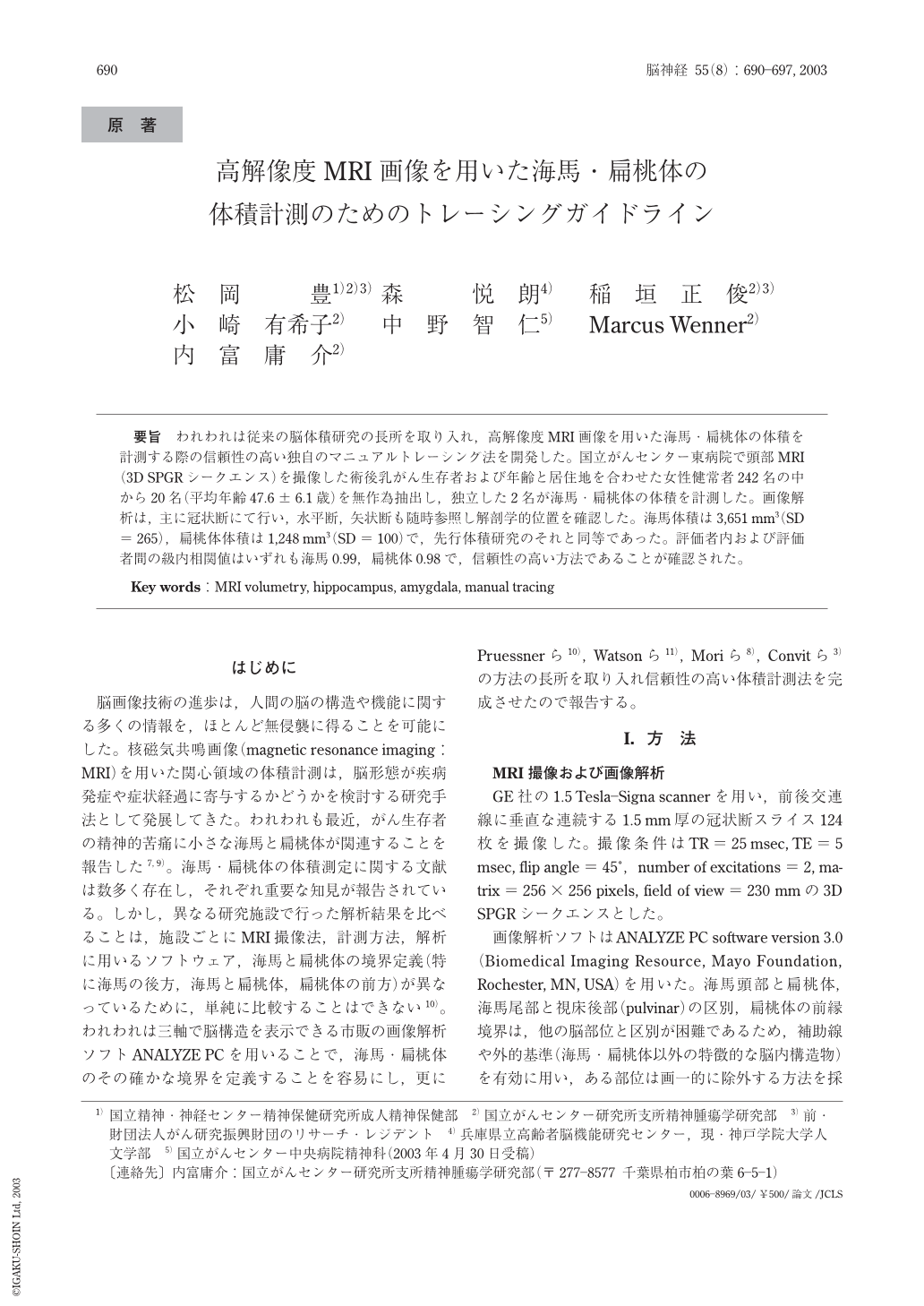
要旨 われわれは従来の脳体積研究の長所を取り入れ,高解像度MRI画像を用いた海馬扁桃体の体積を計測する際の信頼性の高い独自のマニュアルトレーシング法を開発した。国立がんセンター東病院で頭部MRI(3D SPGRシークエンス)を撮像した術後乳がん生存者および年齢と居住地を合わせた女性健常者242名の中から20名(平均年齢47.6±6.1歳)を無作為抽出し,独立した2名が海馬扁桃体の体積を計測した。画像解析は,主に冠状断にて行い,水平断,矢状断も随時参照し解剖学的位置を確認した。海馬体積は3,651mm3(SD=265),桃体体積は1,248mm3(SD=100)で,先行体積研究のそれと同等であった。評価者内および評価者間の級内相関値はいずれも海馬0.99,扁桃体0.98で,信頼性の高い方法であることが確認された。
Both the hippocampus and amygdala are frequently targeted by researchers and clinicians for volumetric analysis based on magnetic resonance imaging(MRI). However, different data acquisition, analysis software and anatomical boundaries have in the past made it difficult to compare results of MRI studies from different laboratories. A reliable original segmentation protocol for volumetry of the amygdala and hippocampus on three-dimensional MRI was established by including merits of previous volumetric studies. Two skilled raters measured randomly selected 20 subjects from 242 subjects recently scanned in our institution(mean age 47.6±6.1 years). Data acquisition was performed with a three-dimensional spoiled gradient echo technique. Volumetric analysis was performed manually using three-dimensional software that allowed coronal, sagittal, and transverse images at any time. Both intra- and inter-rater reliability(intraclass correlation coefficients)yielded 0.99 for the hippocampus and 0.98 for the amygdala. It was confirmed that this segmentation protocol was highly reliable.

Copyright © 2003, Igaku-Shoin Ltd. All rights reserved.


