Japanese
English
- 有料閲覧
- Abstract 文献概要
- 1ページ目 Look Inside
- 参考文献 Reference
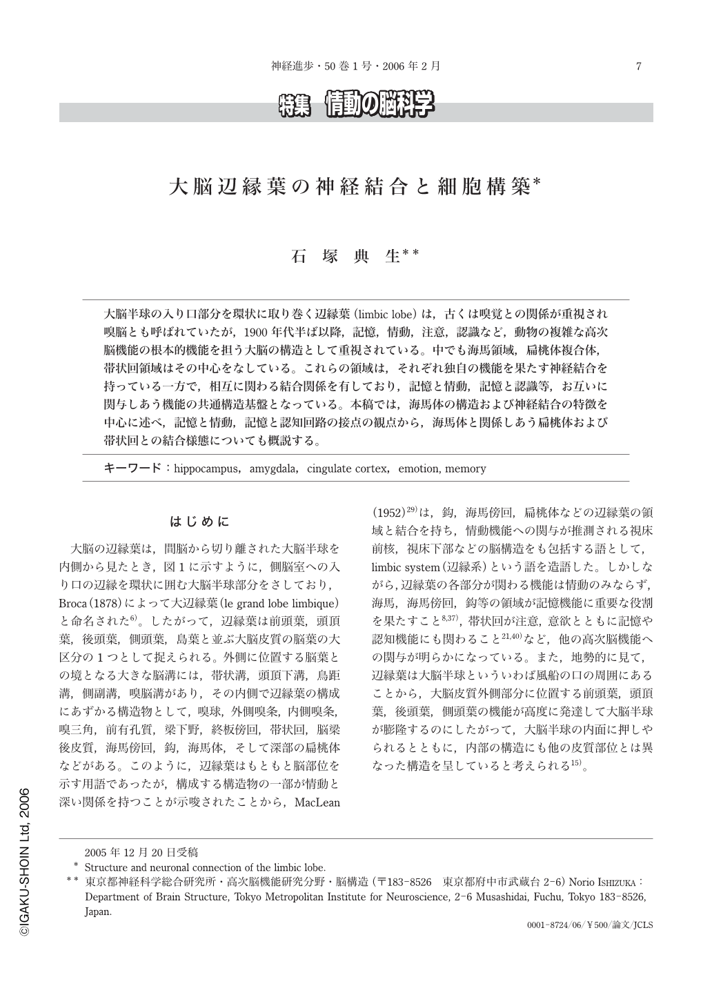
大脳半球の入り口部分を環状に取り巻く辺縁葉(limbic lobe)は,古くは嗅覚との関係が重視され嗅脳とも呼ばれていたが,1900年代半ば以降,記憶,情動,注意,認識など,動物の複雑な高次脳機能の根本的機能を担う大脳の構造として重視されている。中でも海馬領域,扁桃体複合体,帯状回領域はその中心をなしている。これらの領域は,それぞれ独自の機能を果たす神経結合を持っている一方で,相互に関わる結合関係を有しており,記憶と情動,記憶と認識等,お互いに関与しあう機能の共通構造基盤となっている。本稿では,海馬体の構造および神経結合の特徴を中心に述べ,記憶と情動,記憶と認知回路の接点の観点から,海馬体と関係しあう扁桃体および帯状回との結合様態についても概説する。
The limbic lobe is located at the most medial portion(limbus)of the cerebral hemisphere, like as a band surrounding the orifice into the lateral ventricle. The limbic lobe is mainly consisted of the hippocampal formation, the amygdaloid complex and the cingulate cortices. These areas are concerned with the basic brain higher functions, such as emotion, memory, attention, cognition, and so on. Each area has the specific structural organization and the fiber connections executing the specific function. The circuit of memory, so called Papez circuit, is consisted of the hippocampal formation, mammillary body, anterior and midline thalamic nuclei, posterior cingulate cortex, and the retrohippocampal cortices. On the other hand, the circuit of the emotion, so called Yakovlev circuit, is consisted of the amygdala, the mediodorsal thalamic nucleus, anterior cingulate cortex and the orbitofrontal cortex. Although memory and emotion are processed on the independent circuit, there are also several structures where the fibers from the two circuits meet together, such as the nucleus accumbens, the entorhinal cortex, and the hypothalamic area. In this paper, we first describe the specific structure and the fiber connection of the hippocampal formation, and subsequently those of the amygdala and the cingulate cortex in relation to the hippocampal formation.
The hippocampal formation is consisted of the dentate gyrus, the hippocampus proper(CA1 and CA3)and the subiculum. The hippocampal subcortical efferent pathways are originated from the subiculum. The cells locating in the deepest layer of the subiculum give rise to projections to the anteroventral thalamic nucleus(AV), those in the vertically middle layer of the subiculum give rise to projections to the medial nucleus of the mammillary body(MMB), and those in the superficial layer of the subiculum give rise to projections to the nucleus accumbens(ventral striatum). Considering from the view point of output connections of the principle cells of each subfield, we have proposed a hypothesis that the subiculum corresponds to layers Ⅴ and Ⅵ of the isocotex(infragranular layer), a nd also that the dentate gyrus and the hippocampus proper may correspond to layers Ⅱ and Ⅲ(supragranular layer).
The subiculum gives rise to the cortical projections to the entorhinal cortex(MEA, LEA), presubiculum(PRE), parasubiculum(PARA), the retrosplenial granular cortex(RSG), the perirhinal cortex and the postrhinal cortex. Cells of origin of the projections to the MEA, LEA, PARA, PRE, RSG were consistently observed throughout the entire septotemporal extent of the subiculum. In the transverse plane,the cortical projection cells to the MEA, PARA, PRE, RSG were located in the region of the MMB cells, which is in the vertically mid portion of the subicular pyramidal cell layer avoiding the deepest and most superficial portions of the layer. The cells giving rise to the projections to the LEA were located in the superficial portion of the subicular pyramidal cell layer in which cells projecting to the nucleus accumbens were densely located. The septal subiculum gives rise to the projections to the septal parts of RSG, PARA and PRE, and to the lateral(close to the rhinal fissure)part of MEA and LEA, while the temporal subiculum gives rise to the projections to the reverse sides of the areas. Cells located in the proximal part of each region innervate the proximal parts of the PRE and RSG, and the region close to the border between MEA and LEA, while those in the distal part of the region gives rose to the projections to the reverse sides of the areas.
Basal nucleus and accessory basal nucleus of the amygdala have reciprocal connections with the LEA, the subiculum, CA1, perirhinal cortex and the anterior cigulate cortex. Thus, in the future, the mutual connections among the hippocampal formation, the amygdala and the cingulate cortex have to be elucidated.

Copyright © 2006, Igaku-Shoin Ltd. All rights reserved.


