Japanese
English
- 有料閲覧
- Abstract 文献概要
- 1ページ目 Look Inside
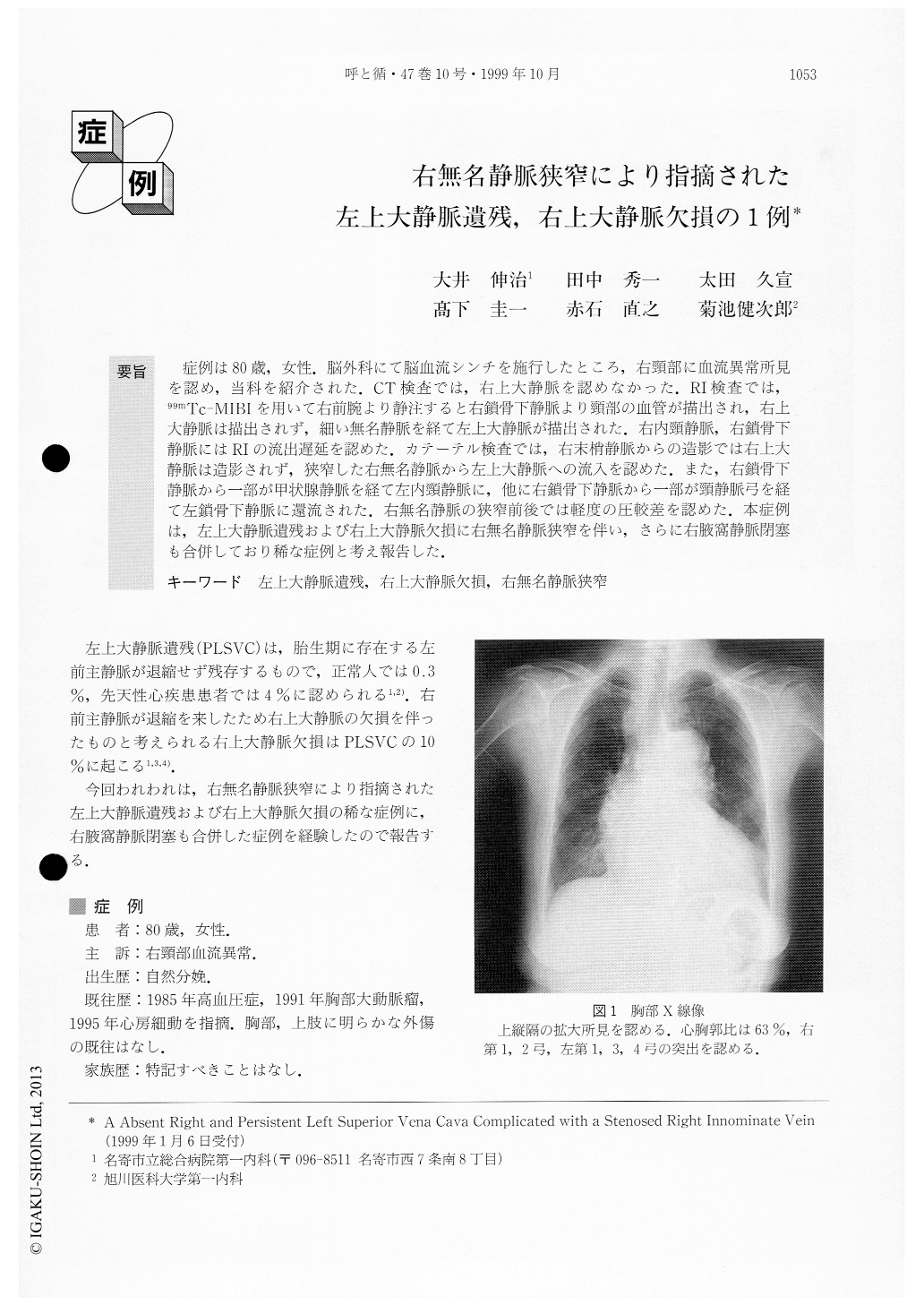
症例は80歳,女性.脳外科にて脳血流シンチを施行したところ、右頸部に血流異常所見を認め,当科を紹介された.CT検査では,右上大静脈を認めなかった.RI検査では,99mTc-MIBIを用いて右前腕より静注すると右鎖骨下静脈より頸部の血管が描出され,右上大静脈は描出されず,細い無名静脈を経て左上大静脈が描出された.右内頸静脈,右鎖骨下静脈にはRIの流出遅延を認めた.カテーテル検査では,右末梢静脈からの造影では右上大静脈は造影されず,狭窄した右無名静脈から左上大静脈への流入を認めた.また,右鎖骨下静脈から一部が甲状腺静脈を経て左内頸静脈に,他に右鎖骨下静脈から一部が頸静脈弓を経て左鎖骨下静脈に還流された.右無名静脈の狭窄前後では軽度の圧較差を認めた.本症例は,左上大静脈遺残および右上大静脈欠損に右無名静脈狭窄を伴い,さらに右腋窩静脈閉塞も合併しており稀な症例と考え報告した.
We reported here a very rare case of a patient with an absent right and persistent left superior vena cava with innominate vein stenosis. The patient was an 80-year-old woman who had undergone injection of radio-isotopes from the right radial vein because of cerebral artery sclerosis. Radioisotope examination revealed an abnormality of a right innominate vein, so we were consulted about the woman by a neurosurgeon.
A venous angiocardiogram was performed from the right radial vein. The contrast material was seen toenter the stenosed right innominate vein, pass the left superior vena cava and reach the right atrium only by way of the left superior vena cava. The right superior vena cava was not visualized. Furthermore, the contrast material showed that the right internal jugular vein entered a part of the right subclavian vein and passed on to the right thyroid vein to reach the left thyroid vein and the left jugular vein. The right external jugular vein entered a part of the right subclavian vein and passed to the jugular venous arch to reach the left external jugu-lar vein and the left subclavian vein.
To our knowledge, this is the first case of an absent right and a persistent left superior vena cava compli-cated with a stenosed right innominate vein.

Copyright © 1999, Igaku-Shoin Ltd. All rights reserved.


