Japanese
English
- 有料閲覧
- Abstract 文献概要
- 1ページ目 Look Inside
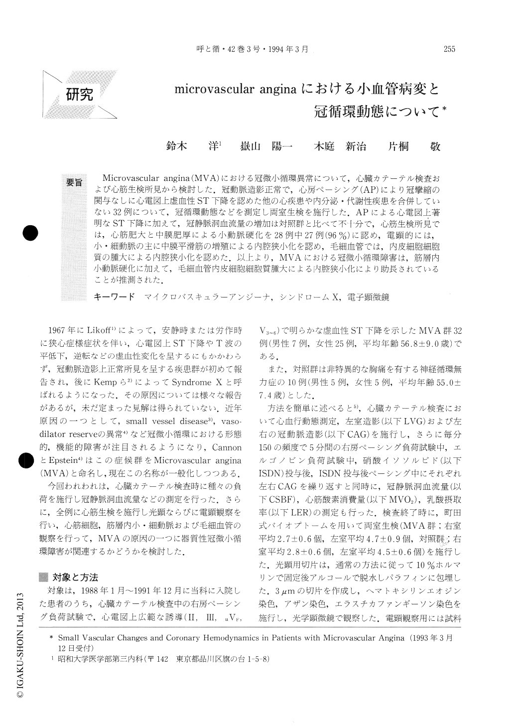
Microvascular angina(MVA)における冠微小循環異常について,心臓カテーテル検査および心筋生検所見から検討した.冠動脈造影正常で,心房ペーシング(AP)により冠攣縮の関与なしに心電図上虚血性ST下降を認めた他の心疾患や内分泌・代謝性疾患を合併していない32例について,冠循環動態などを測定し両室生検を施行した.APによる心電図上著明なST下降に加えて,冠静脈洞血流量の増加は対照群と比べて不十分で,心筋生検所見では,心筋肥大と中膜肥厚による小動脈硬化を28例中27例(96%)に認め,電顕的には,小・細動脈の主に中膜平滑筋の増殖による内腔狭小化を認め,毛細血管では,内皮細胞細胞質の腫大による内腔狭小化を認めた.以上より,MVAにおける冠微小循環障害は,筋層内小動脈硬化に加えて,毛細血管内皮細胞細胞質腫大による内腔狭小化により助長されていることが推測された.
We studied ultrastructural changes of cardiac myocytes and small blood vessels obtained by endo-myocardial biopsy in patients with microvascular an-gina (MVA).
Thirty-two patients with ST segment depression during atrial pacing with normal coronary angiograms and without any provocated spasm were examined. During pacing, they were checked for chest pain and ECG changes by measuring coronary sinus blood flow (CSBF), myocardial oxygen consumption (MVO2) etc. and coronary angiographies were repeated. Myocardial tissues, biopsied from both ventricles were observed with a light and with an electron microscope.

Copyright © 1994, Igaku-Shoin Ltd. All rights reserved.


