Japanese
English
- 有料閲覧
- Abstract 文献概要
- 1ページ目 Look Inside
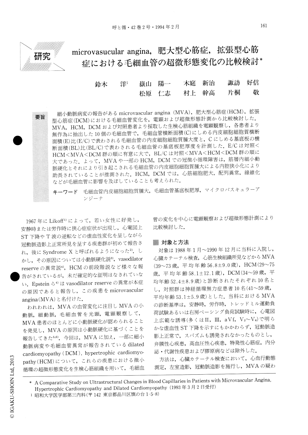
細小動脈病変の報告があるmicrovascular angina(MVA),肥大型心筋症(HCM),拡張型心筋症(DCM)における毛細血管変化を,電顕および超微形態計測から比較検討した.MVA,HCM,DCMおよび対照患者より採取した生検心筋組織を電顕観察し,各患者より無作為に抽出した10個の毛細血管で,毛細血管横断面積(C)にしめる内皮細胞細胞質横断面積(E)比(E/C)で表わされる毛細血管の内皮細胞細胞質腫大度と,Cにしめる基底板の横断面積(BL)比(BL/C)で表わされる毛細血管の基底板肥厚度を計測した.E/Cは対照<HCM<MVA<DCM群の順に有意に大で,BL/Cは対照<MVA<HCM<DCM群の順に大であった.よって,MVAや一部のHCM,DCMでの冠微小循環障害は,筋層内細小動脈硬化とそれにより引き起こされる毛細血管の内皮細胞細胞質腫大による内腔狭小化により助長されていることが推測された.HCM,DCMでは,心筋細胞肥大,配列異常,線維化などが毛細血管に影響を及ぼしていることも考えられた.
There are some reports on capillary abnormalities accompanied by intramural small arterial lesions in microvascular angina (MVA), hypertrophic car-diomyopathy (HCM) and dilated cardiomyopathy (DCM). Using electron microscopy and ultrastructural morphometry, we performed comparative studies on ultrastructural changes in capillaries taken from biopsy samples in MVA, HCM and DCM. Endomyocardial biopsy from both ventricles was carried out during cardiac catheterization in 10 cases of MVA, HCM and DCM and 10 control patients with neurocirculatory asthenia. Then, we examined the specimens with elec-tron microscopy.

Copyright © 1994, Igaku-Shoin Ltd. All rights reserved.


