Japanese
English
- 有料閲覧
- Abstract 文献概要
- 1ページ目 Look Inside
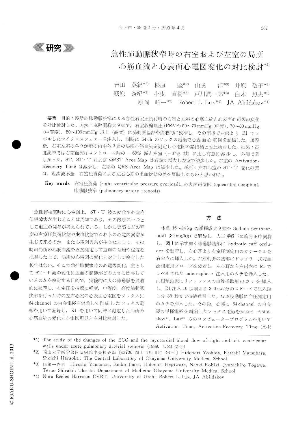
目的:段階的肺動脈狭窄による急性右室圧負荷時の右室と左室の心筋血流と心表面心電図の変化を対比検討した。方法:麻酔開胸犬9頭で,右室収縮期圧(PRVP)50〜70 mmHg(軽度),70〜80 mmHg(中等度),80〜100 mmHg以上(高度)に肺動脈基部を段階的に狭窄し,その前後で左房よりRIでラベルしたマイクロスフェアーを注入し,同時に64chのソックス電極で心表面心電図を記録した。屠殺後,右室左室の各9か所の内中外3層の局所心筋血流を測定し心電図の諸指標と対比検討した。結果:高度狭窄では右室血流はコントロール時の−63%減と左室(−37%減)に比し有意に減少し,外層で著しかった。ST, ST・TおよびQRST Area Mapは右室で増大し左窒で減少した。右室のActivation-Recovery Timeは減少し,左室のQRS Area Mapは減少した。総括;左右心室のST・T変化の差は,冠灌流不全,右室圧負荷による左右心筋の虚血状態の差を反映したものと思われた。
For studying the effect of regional myocardial blood flow changes on the epicardial ECG of right and left ventricular walls under acute right ventri-cular pressure overload, we mapped the epicardial using 64-channel sock electrodes, and estimated the myocardial blood flow with radioactive microshe-res. In 9 anesthetized open-chest dogs, the main pulmonary artery was gradedly contricted to the level of mild (peak RV pressure: PRVP, 50~70 mmHg), moderate (PRVP, 70~80 mmHg) and severe stenosis (PRVP, over 80mmHg). Labeled micro-spheres were injected into the left atrium before and after the PA constriction, and the epicardial ECGs were recorded continuously. After the comp-letion of the experiment, 9 areas of each right and left ventricular wall were excised. The myocardium was divided into three layers and the flow data were compared to the changes of ECG parameters. In the cases where there was severe PA stenosis, the right ventricular myocardial blood flow decreased to asignificantly greater degree (63% reduction from the control), especially in the subepicardial layer, than the flow in the left ventricle (37% reduction from the control). ST potential, STT and QRST Area Map increased in the right ventricle but decreased in the left ventricle. Activation Recovery Time of the right ventricle decreased due to the severe is-chemia of the right ventricle. The value of QRSArea Map of the left ventricle decreased signifi-cantly in parallel with the decrease in cardiac out-put. The data suggested that the right ventricle is exposed to more severe ischemia than the left vent-ricle. This is due to lowered coronary perfusion pressure in addition to the direct effect of the right ventricular pressure overload.

Copyright © 1990, Igaku-Shoin Ltd. All rights reserved.


